Japanese
English
- 有料閲覧
PPV購入案内
1,320円 (1,200円+税10%) こちらは、149ページから155ページの文献です。購入には医書.jp本会員登録が必要です。
会員登録完了後に改めて上記「購入サイトへ」を選択してください。
または「購入サイトへ」選択後に「購入ログイン」、「新規会員登録」の順に進んでください。
会員登録について詳しくはをご確認ください。
- Abstract 文献概要
- 1ページ目 Look Inside
- 参考文献 Reference
Ⅰ.はじめに
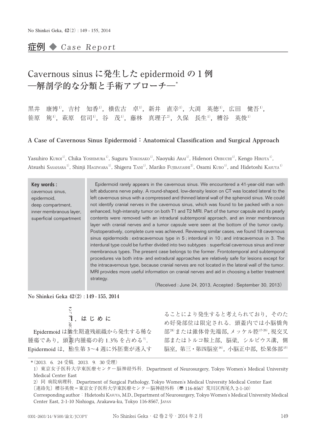
Epidermoidは胎生期遺残組織から発生する稀な腫瘍であり,頭蓋内腫瘍の約1.3%を占める7).Epidermoidは,胎生第3~4週に外胚葉が迷入することにより発生すると考えられており,そのため好発部位は限定される.頭蓋内では小脳橋角部28)または錐体骨先端部,メッケル腔17-20),視交叉部またはトルコ鞍上部,脳梁,シルビウス溝,側脳室,第三・第四脳室16),小脳正中部,松果体部15)に好発する.また,脊髄腔内では腰仙部に好発する.一方,海綿静脈洞近傍に発生するepidermoidは非常に稀であり,現在に至るまで20例程度の報告があるのみである4-6,8-12,14,20-23,26,29).今回われわれは左外転神経麻痺で発症したepidermoidを経験したので,文献的考察を交えて報告する.
Epidermoid rarely appears in the cavernous sinus. We encountered a 41-year-old man with left abducens nerve palsy. A round-shaped, low-density lesion on CT was located lateral to the left cavernous sinus with a compressed and thinned lateral wall of the sphenoid sinus. We could not identify cranial nerves in the cavernous sinus, which was found to be packed with a non-enhanced, high-intensity tumor on both T1 and T2 MRI. Part of the tumor capsule and its pearly contents were removed with an intradural subtemporal approach, and an inner membranous layer with cranial nerves and a tumor capsule were seen at the bottom of the tumor cavity. Postoperatively, complete cure was achieved. Reviewing similar cases, we found 18 cavernous sinus epidermoids:extracavernous type in 5;interdural in 10;and intracavernous in 3. The interdural type could be further divided into two subtypes:superficial cavernous sinus and inner membranous types. The present case belongs to the former. Frontotemporal and subtemporal procedures via both intra- and extradural approaches are relatively safe for lesions except for the intracavernous type, because cranial nerves are not located in the lateral wall of the tumor. MRI provides more useful information on cranial nerves and aid in choosing a better treatment strategy.
Copyright © 2014, Igaku-Shoin Ltd. All rights reserved.