Japanese
English
- 有料閲覧
PPV購入案内
1,320円 (1,200円+税10%) こちらは、149ページから155ページの文献です。購入には医書.jp本会員登録が必要です。
会員登録完了後に改めて上記「購入サイトへ」を選択してください。
または「購入サイトへ」選択後に「購入ログイン」、「新規会員登録」の順に進んでください。
会員登録について詳しくはをご確認ください。
- Abstract 文献概要
- 1ページ目 Look Inside
- 参考文献 Reference
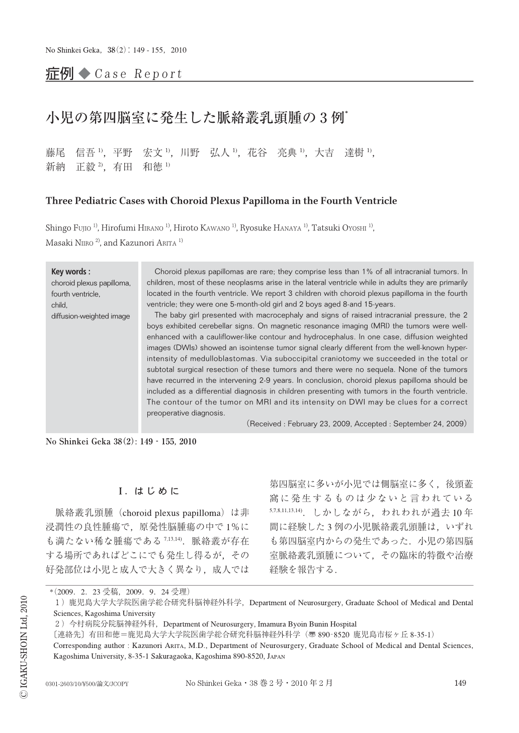
Ⅰ.はじめに
脈絡叢乳頭腫(choroid plexus papilloma)は非浸潤性の良性腫瘍で,原発性脳腫瘍の中で1%にも満たない稀な腫瘍である7,13,14).脈絡叢が存在する場所であればどこにでも発生し得るが,その好発部位は小児と成人で大きく異なり,成人では第四脳室に多いが小児では側脳室に多く,後頭蓋窩に発生するものは少ないと言われている5,7,8,11,13,14).しかしながら,われわれが過去10年間に経験した3例の小児脈絡叢乳頭腫は,いずれも第四脳室内からの発生であった.小児の第四脳室脈絡叢乳頭腫について,その臨床的特徴や治療経験を報告する.
Choroid plexus papillomas are rare; they comprise less than 1% of all intracranial tumors. In children, most of these neoplasms arise in the lateral ventricle while in adults they are primarily located in the fourth ventricle. We report 3 children with choroid plexus papilloma in the fourth ventricle; they were one 5-month-old girl and 2 boys aged 8-and 15-years.
The baby girl presented with macrocephaly and signs of raised intracranial pressure, the 2 boys exhibited cerebellar signs. On magnetic resonance imaging (MRI) the tumors were well-enhanced with a cauliflower-like contour and hydrocephalus. In one case, diffusion weighted images (DWIs) showed an isointense tumor signal clearly different from the well-known hyper-intensity of medulloblastomas. Via suboccipital craniotomy we succeeded in the total or subtotal surgical resection of these tumors and there were no sequela. None of the tumors have recurred in the intervening 2-9 years. In conclusion, choroid plexus papilloma should be included as a differential diagnosis in children presenting with tumors in the fourth ventricle. The contour of the tumor on MRI and its intensity on DWI may be clues for a correct preoperative diagnosis.
Copyright © 2010, Igaku-Shoin Ltd. All rights reserved.