Japanese
English
- 有料閲覧
PPV購入案内
1,320円 (1,200円+税10%) こちらは、495ページから501ページの文献です。購入には医書.jp本会員登録が必要です。
会員登録完了後に改めて上記「購入サイトへ」を選択してください。
または「購入サイトへ」選択後に「購入ログイン」、「新規会員登録」の順に進んでください。
会員登録について詳しくはをご確認ください。
- Abstract 文献概要
- 1ページ目 Look Inside
- 参考文献 Reference
Ⅰ.はじめに
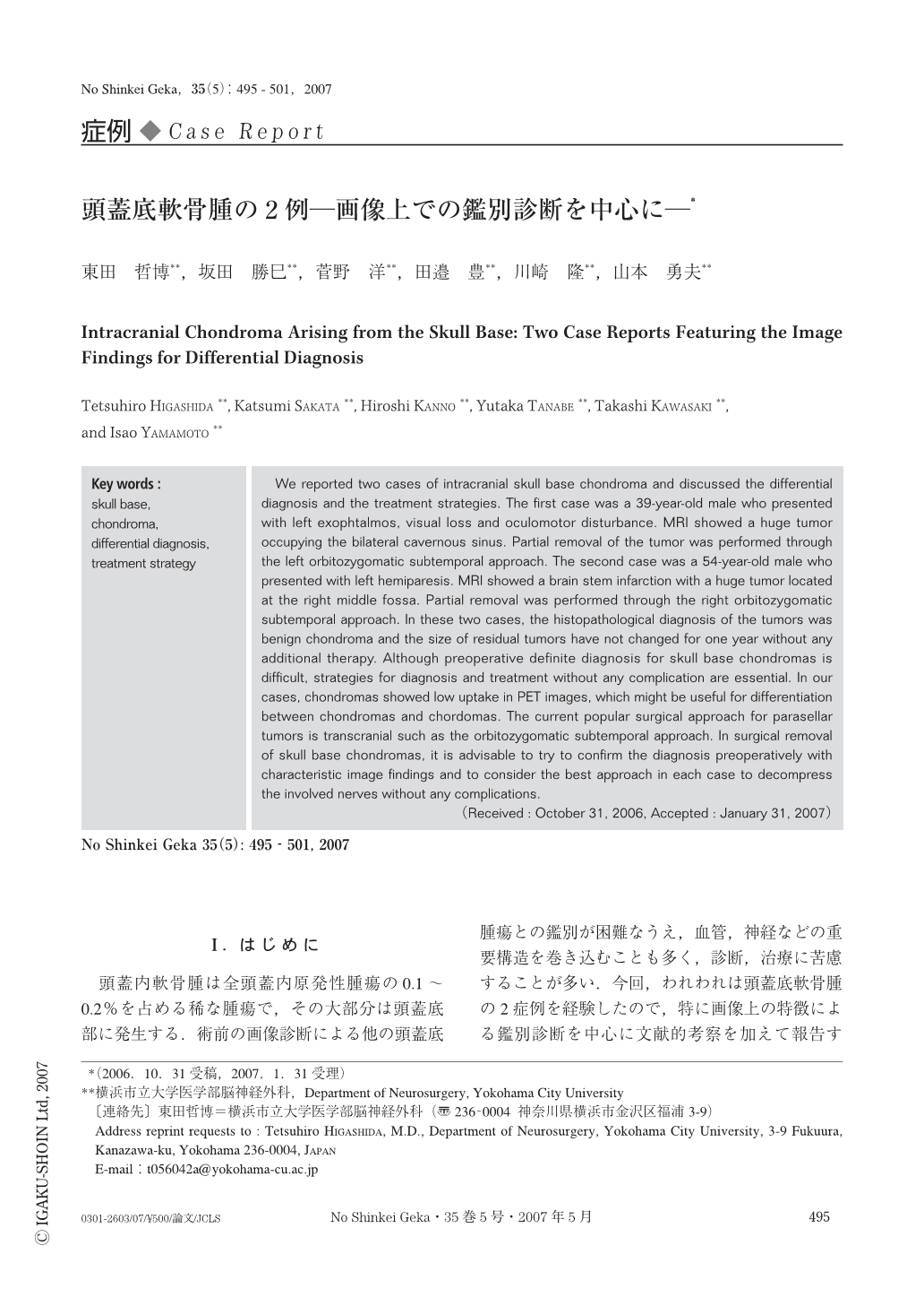
頭蓋内軟骨腫は全頭蓋内原発性腫瘍の0.1~0.2%を占める稀な腫瘍で,その大部分は頭蓋底部に発生する.術前の画像診断による他の頭蓋底腫瘍との鑑別が困難なうえ,血管,神経などの重要構造を巻き込むことも多く,診断,治療に苦慮することが多い.今回,われわれは頭蓋底軟骨腫の2症例を経験したので,特に画像上の特徴による鑑別診断を中心に文献的考察を加えて報告する.
We reported two cases of intracranial skull base chondroma and discussed the differential diagnosis and the treatment strategies. The first case was a 39-year-old male who presented with left exophtalmos, visual loss and oculomotor disturbance. MRI showed a huge tumor occupying the bilateral cavernous sinus. Partial removal of the tumor was performed through the left orbitozygomatic subtemporal approach. The second case was a 54-year-old male who presented with left hemiparesis. MRI showed a brain stem infarction with a huge tumor located at the right middle fossa. Partial removal was performed through the right orbitozygomatic subtemporal approach. In these two cases, the histopathological diagnosis of the tumors was benign chondroma and the size of residual tumors have not changed for one year without any additional therapy. Although preoperative definite diagnosis for skull base chondromas is difficult, strategies for diagnosis and treatment without any complication are essential. In our cases, chondromas showed low uptake in PET images, which might be useful for differentiation between chondromas and chordomas. The current popular surgical approach for parasellar tumors is transcranial such as the orbitozygomatic subtemporal approach. In surgical removal of skull base chondromas, it is advisable to try to confirm the diagnosis preoperatively with characteristic image findings and to consider the best approach in each case to decompress the involved nerves without any complications.
Copyright © 2007, Igaku-Shoin Ltd. All rights reserved.