Japanese
English
- 有料閲覧
PPV購入案内
1,320円 (1,200円+税10%) こちらは、861ページから868ページの文献です。購入には医書.jp本会員登録が必要です。
会員登録完了後に改めて上記「購入サイトへ」を選択してください。
または「購入サイトへ」選択後に「購入ログイン」、「新規会員登録」の順に進んでください。
会員登録について詳しくはをご確認ください。
- Abstract 文献概要
- 1ページ目 Look Inside
- 参考文献 Reference
はじめに
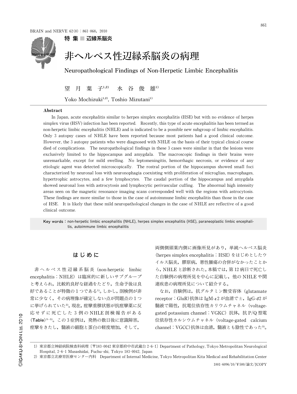
非ヘルペス性辺縁系脳炎(non-herpetic limbic encephalitis:NHLE)は臨床的に新しいサブグループと考えられ,比較的良好な経過をたどり,生命予後は良好であることが特徴の1つである1)。しかし,剖検例が非常に少なく,その病理像が確定しない点が問題点の1つに挙げられていた2)。現在,痙攣重積状態が抗痙攣薬に反応せずに死亡した3例のNHLE剖検報告がある(Table)3-5)。この3症例は,発熱の数日後に意識障害,痙攣をきたし,髄液の細胞と蛋白の軽度増加,そして,両側側頭葉内側に画像所見があり,単純ヘルペス脳炎(herpes simplex encephalitis:HSE)をはじめとしたウイルス脳炎,膠原病,悪性腫瘍の合併がなかったことから,NHLEと診断された。本稿では,第12病日で死亡した自験例の病理所見を中心に記載し,他のNHLEや関連疾患の病理所見について紹介する。
なお,自験例は,抗グルタミン酸受容体(glutamate receptor:GluR)抗体はIgM-ε2が血清で±,IgG-δ2が髄液で陽性,抗電位依存性カリウムチャネル(voltage-gated potassium channel:VGKC)抗体,抗P/Q型電位依存性カルシウムチャネル(voltage-gated calcium channel:VGCC)抗体は血清,髄液とも陰性であった3)。
Abstract
In Japan,acute encephalitis similar to herpes simplex encephalitis (HSE) but with no evidence of herpes simplex virus (HSV) infection has been reported. Recently,this type of acute encephalitis has been termed as non-herpetic limbic encephalitis (NHLE) and is indicated to be a possible new subgroup of limbic encephalitis. Only 3 autopsy cases of NHLE have been reported because most patients had a good clinical outcome. However,the 3 autopsy patients who were diagnosed with NHLE on the basis of their typical clinical course died of complications. The neuropathological findings in these 3 cases were similar in that the lesions were exclusively limited to the hippocampus and amygdala. The macroscopic findings in their brains were unremarkable,except for mild swelling. No leptomeningitis,hemorrhagic necrosis,or evidence of any etiologic agent was detected microscopically. The rostral portion of the hippocampus showed small foci characterized by neuronal loss with neuronophagia coexisting with proliferation of microglias,macrophages,hypertrophic astrocytes,and a few lymphocytes. The caudal portion of the hippocampus and amygdala showed neuronal loss with astrocytosis and lymphocytic perivascular cuffing. The abnormal high intensity areas seen on the magnetic resonance imaging scans corresponded well with the regions with astrocytosis. These findings are more similar to those in the case of autoimmune limbic encephalitis than those in the case of HSE. It is likely that these mild neuropathological changes in the case of NHLE are reflective of a good clinical outcome.
Copyright © 2010, Igaku-Shoin Ltd. All rights reserved.