Japanese
English
- 有料閲覧
PPV購入案内
1,320円 (1,200円+税10%) こちらは、1061ページから1064ページの文献です。購入には医書.jp本会員登録が必要です。
会員登録完了後に改めて上記「購入サイトへ」を選択してください。
または「購入サイトへ」選択後に「購入ログイン」、「新規会員登録」の順に進んでください。
会員登録について詳しくはをご確認ください。
- Abstract 文献概要
- 1ページ目 Look Inside
- 参考文献 Reference
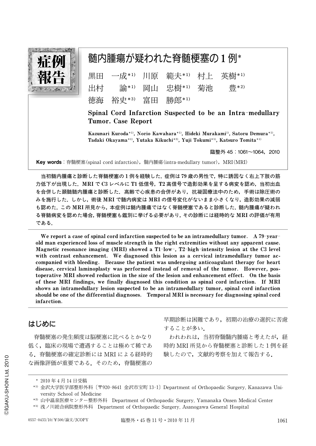
当初髄内腫瘍と診断した脊髄梗塞の1例を経験した.症例は79歳の男性で,特に誘因なく右上下肢の筋力低下が出現した.MRIでC3レベルにT1低信号,T2高信号で造影効果を呈する病変を認め,当初出血を合併した頚髄髄内腫瘍と診断した.高齢で心疾患の合併があり,抗凝固療法中のため,手術は除圧術のみを施行した.しかし,術後MRIで髄内病変はMRIの信号変化がないまま小さくなり,造影効果の減弱も認めた.このMRI所見から,本症例は髄内腫瘍ではなく脊髄梗塞であると診断した.髄内腫瘍が疑われる脊髄病変を認めた場合,脊髄梗塞も鑑別に挙げる必要があり,その診断には経時的なMRIの評価が有用である.
We report a case of spinal cord infarction suspected to be an intramedullary tumor. A 79-year-old man experienced loss of muscle strength in the right extremities without any apparent cause. Magnetic resonance imaging (MRI) showed a T1-low-, T2-high-intensity lesion at the C3 level with contrast enhancement. We diagnosed this lesion as a cervical intramedullary tumor accompanied with bleeding. Because the patient was undergoing anticoagulant therapy for heart disease, cervical laminoplasty was performed instead of removal of the tumor. However, postoperative MRI showed reduction in the size of the lesion and enhancement effect. On the basis of these MRI findings, we finally diagnosed this condition as spinal cord infarction. If MRI shows an intramedullary lesion suspected to be an intramedullary tumor, spinal cord infarction should be one of the differential diagnoses. Temporal MRI is necessary for diagnosing spinal cord infarction.
Copyright © 2010, Igaku-Shoin Ltd. All rights reserved.