Japanese
English
- 有料閲覧
PPV購入案内
1,320円 (1,200円+税10%) こちらは、77ページから81ページの文献です。購入には医書.jp本会員登録が必要です。
会員登録完了後に改めて上記「購入サイトへ」を選択してください。
または「購入サイトへ」選択後に「購入ログイン」、「新規会員登録」の順に進んでください。
会員登録について詳しくはをご確認ください。
- Abstract 文献概要
- 1ページ目 Look Inside
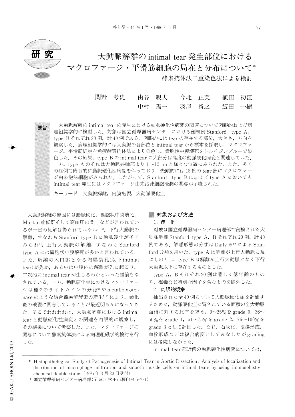
大動脈解離のintimal tearの発生における動脈硬化性病変の関連について肉眼的および病理組織学的に検討した.対象は国立循環器病センターにおける剖検例Stanford type A,type Bそれぞれ20例,計40例である.肉眼的にはtearの存在する部位,大きさ,方向を観察した.病理組織学的には大動脈の各部位とintimal tearから標本を採取し,マクロファージ,平滑筋細胞を免疫酵素抗体法により染色し,嚢胞性中膜壊死をトルイジンブルーで染色した.その結果,type Bのintimal tearの大部分は高度の動脈硬化病変と関連していた.一方,type Aのそれは大動脈弁輪部より1〜12cmと様々な位置にみられた.また,多くの症例で肉眼的に動脈硬化性病変を伴っており,光顕的には18例のtear部にマクロファージ由来泡沫細胞がみられた.したがって,Stanford type Bに加えてtype Aにおいてもintinnal tear発生にはマクロファージ由来泡沫細胞浸潤の関与が示唆された.
In aortic dissection, we have grossly and histopath-ologically examined intimal tears to disclose the rela-tion between them and atherosclerosis. We studied 20 autopsy cases with both Stanford type A and Stanford type B. The location, width and direction of intimal tears were grossly observed. Serial histological sections were obtained from intimal tears and segments of the aorta. They were stained with enzyme immunoassay to find the distribution of macrophages and smooth muscle cells. Cystic medial necrosis was evaluated with Toluidine blue stain. All cases of type B were associat-ed with severe degree of atherosclerotic lesions. On the other hand, foam cell infiltration, which was derived from macrophages, was observed around the intimal tears in 18 cases of type A. The gross and histologic findings and associated circumstances suggest that foam cell infiltration may play an important role in the occurrence of intimal tear in type A aortic dissection as well as in type B.
Copyright © 1996, Igaku-Shoin Ltd. All rights reserved.