Japanese
English
- 有料閲覧
PPV購入案内
1,320円 (1,200円+税10%) こちらは、43ページから54ページの文献です。購入には医書.jp本会員登録が必要です。
会員登録完了後に改めて上記「購入サイトへ」を選択してください。
または「購入サイトへ」選択後に「購入ログイン」、「新規会員登録」の順に進んでください。
会員登録について詳しくはをご確認ください。
- Abstract 文献概要
- 1ページ目 Look Inside
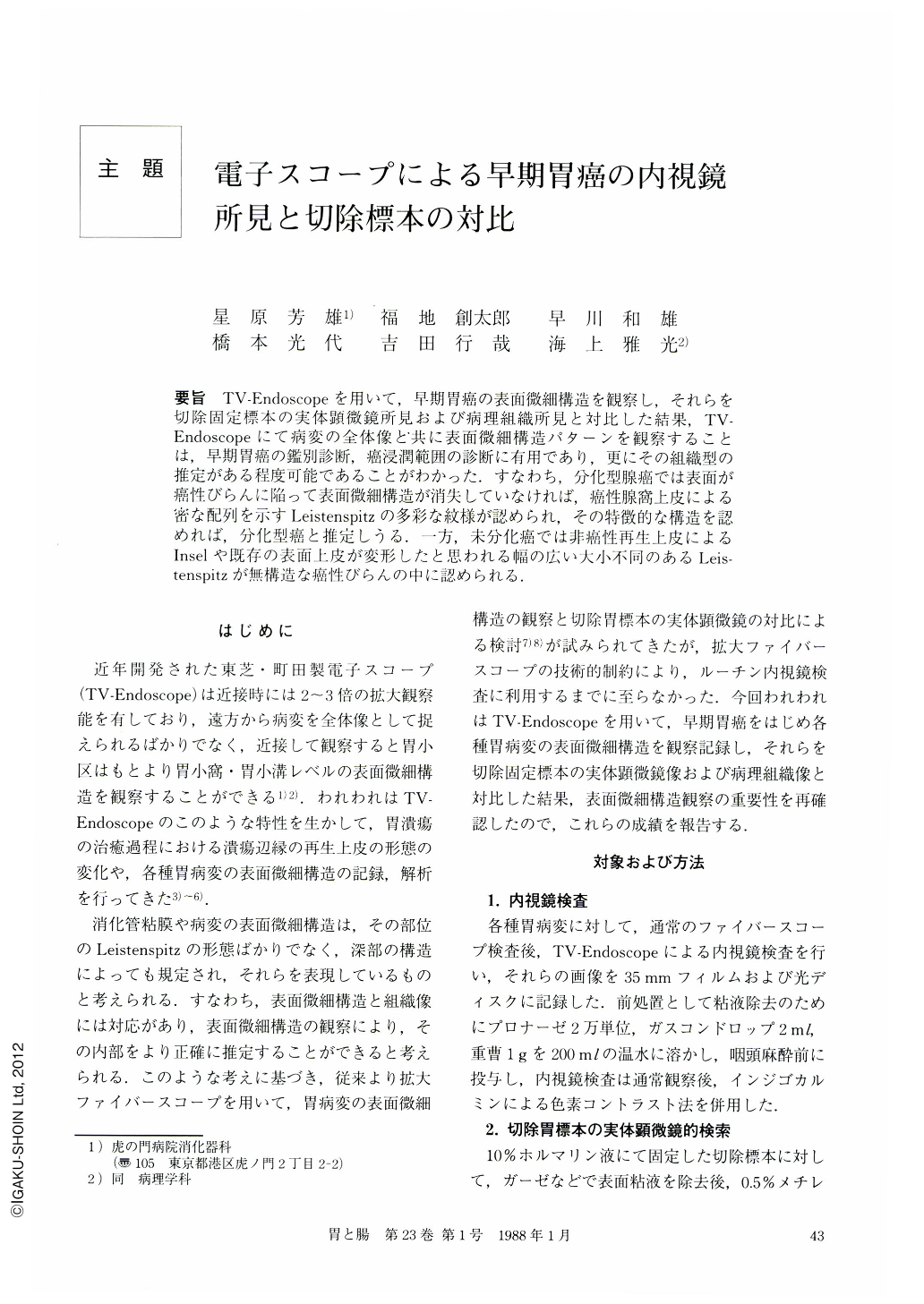
要旨 TV-Endoscopeを用いて,早期胃癌の表面微細構造を観察し,それらを切除固定標本の実体顕微鏡所見および病理組織所見と対比した結果,TV-Endoscopeにて病変の全体像と共に表面微細構造パターンを観察することは,早期胃癌の鑑別診断,癌浸潤範囲の診断に有用であり,更にその組織型の推定がある程度可能であることがわかった.すなわち,分化型腺癌では表面が癌性びらんに陥って表面微細構造が消失していなければ,癌性腺窩上皮による密な配列を示すLeistenspitzの多彩な紋様が認められ,その特徴的な構造を認めれば,分化型癌と推定しうる.一方,未分化癌では非癌性再生上皮によるInselや既存の表面上皮が変形したと思われる幅の広い大小不同のあるLeistenspitzが無構造な癌性びらんの中に認められる.
TV-Endoscopic findings were compared with those obtained by dissecting microscope and histologic examination of surgically resected stomachs. Results are as follows.
1. Minute surface structure seen by TV-Endoscope was confirmed to correspond to those consisted of gastric pits observed by dissecting microscope.
2. Differential diagnosis of early gastric cancer and estimation of histological type are to some degree possible endoscopically by observing whole lesion and such minute surface structure on TV-Endoscope.
3. Dense and diverse Leistenspitz due to carcinomatous tubules is noted in well-differentiated adenocarcinoma unless surface structure is destroyed by carcinomatous erosion. Leistenspitz is most often elongated or linear in shape for elevated type and circular or rod in shape for excavated type.
4. Poorly-differentiated adenocarcinoma has insular foci of regenerative epithelium consisting of wide Leistenspitz in excavated area and relatively large gastric foveolae remaining as a result of destruction of normal gastric mucosa.
5. Possibility of making use of the findings of Leistenspitz in differential diagnosis was discussed by measuring the width of Leistenspitz of both carcinomatous and non-carcinomatous areas by TV-Endoscope and dissecting microscope. There were cases in which the difference in width of Leistenspitz was not significant between carcinomatous and non-carcinomatous areas. Thus, the width of Leistenspitz alone is not useful in differential diagnosis.
Copyright © 1988, Igaku-Shoin Ltd. All rights reserved.