Japanese
English
- 有料閲覧
PPV購入案内
1,320円 (1,200円+税10%) こちらは、675ページから680ページの文献です。購入には医書.jp本会員登録が必要です。
会員登録完了後に改めて上記「購入サイトへ」を選択してください。
または「購入サイトへ」選択後に「購入ログイン」、「新規会員登録」の順に進んでください。
会員登録について詳しくはをご確認ください。
- Abstract 文献概要
- 1ページ目 Look Inside
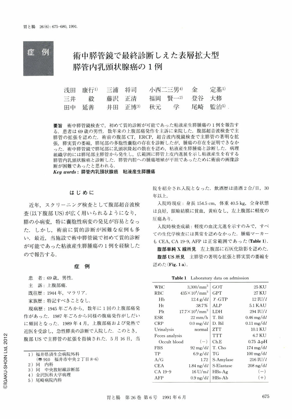
要旨 術中膵管鏡検査で,初めて質的診断が可能であった粘液産生膵腫瘍の1例を報告する.患者は69歳の男性.数年来の上腹部痛発作を主訴に来院した.腹部超音波検査で主膵管の拡張を認めた.術前の腹部CT,ERCP,超音波内視鏡検査で主膵管の著明な拡張,膵実質の萎縮,膵尾部の多胞性囊胞の存在を診断したが,腫瘍の存在を証明できなかった.術中膵管鏡で膵尾部に乳頭状隆起の散在を認め,粘液産生膵腫瘍と診断した.病理組織学的には膵尾部主膵管から発生し,広範囲に膵管上皮内進展を示し粘液産生を有する膵管内乳頭状腺癌と診断した.膵管内腔への腫瘍増殖が平坦であったために術前の画像診断が困難であったと思われる.
A 69-year-old man was admitted to our hospital, because of abdominal pain. Laboratory data on admission was normal except for the elevation of ESR (Table 1). Abdominal ultrasonography (US) demonstrated marked dilatation of the main pancreatic duct (MPD) and atrophy of the pancreas (Fig. 1a). Enhanced CT scan (Fig. 2) and endoscopic ultrasonography (EUS) (Fig. 1b) showed a cystic lesion in the pancreatic tail. Endoscopic retrograde pancreatography (ERP) showed dilatation of the MPD (Fig. 3) in which a lot of mucus was contained. However, examination before the operation demonstrated no neoplastic lesion in the cystic dilatation of the pancreatic duct. By using pancreatoscope during the operation, granular and villous mucosa was revealed in the MPD of the pancreatic tail (Fig. 4). This was diagnosed as mucin producing tumor of the pancreas. Distal pancreatectomy was performed.
Examination of the resected materials revealed granular and low-elevated tumors spreading superficially in the dilated MPD of the pancreatic body and tail (Fig. 6). These tumors were histologically papillary, nonpapillary, vinous, and flat tumors consisting of mucin producing columnar epithelia with nuclear pseudostratification and structural atypia showing severe dysplasia in some parts (Figs. 7, 8). This type of tumor was considered to be intraductal papillary adenocarcinoma (low grade malignancy).
Copyright © 1991, Igaku-Shoin Ltd. All rights reserved.