Japanese
English
- 有料閲覧
- Abstract 文献概要
- 1ページ目 Look Inside
- 参考文献 Reference
- サイト内被引用 Cited by
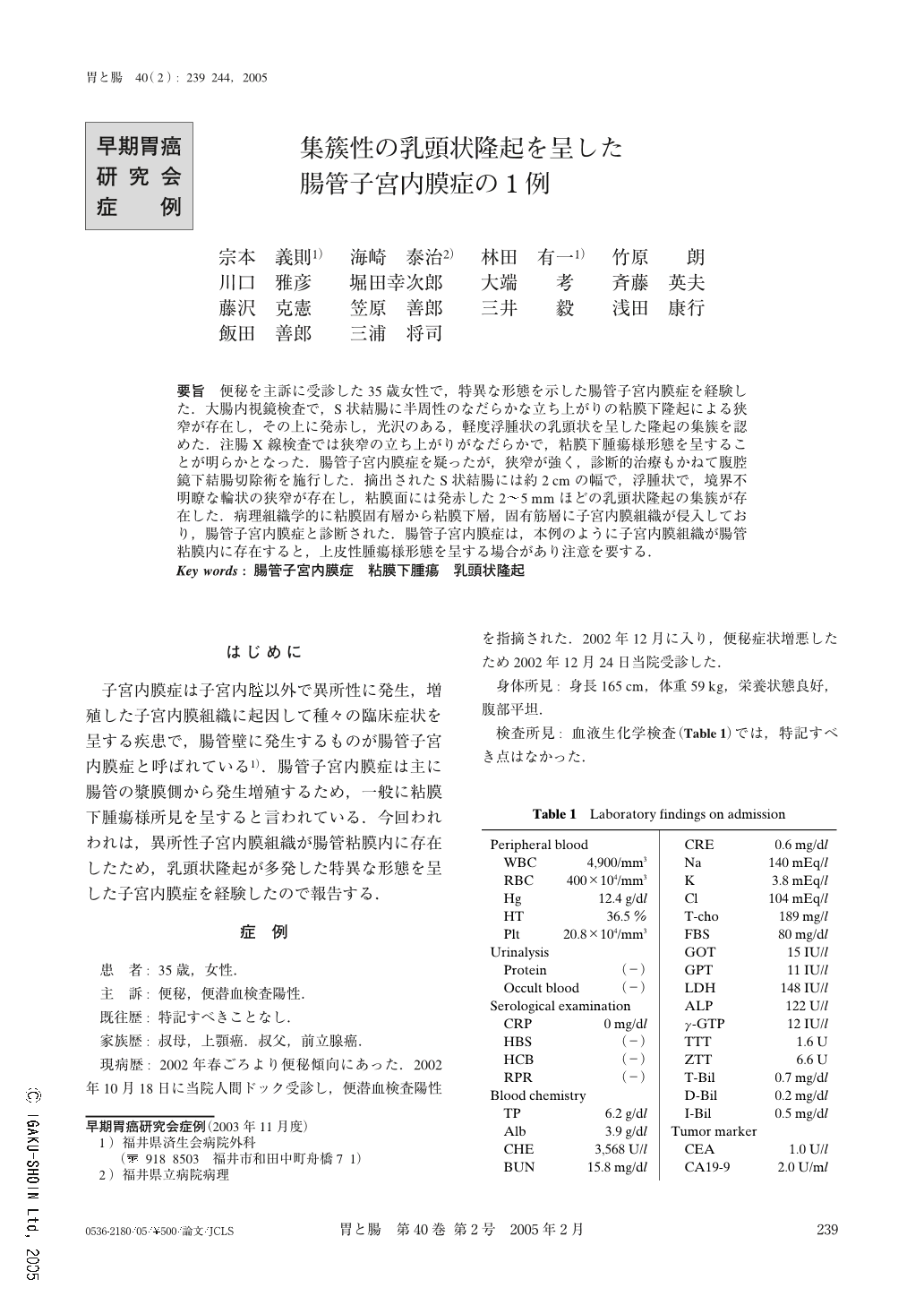
要旨 便秘を主訴に受診した35歳女性で,特異な形態を示した腸管子宮内膜症を経験した.大腸内視鏡検査で,S状結腸に半周性のなだらかな立ち上がりの粘膜下隆起による狭窄が存在し,その上に発赤し,光沢のある,軽度浮腫状の乳頭状を呈した隆起の集簇を認めた.注腸X線検査では狭窄の立ち上がりがなだらかで,粘膜下腫瘍様形態を呈することが明らかとなった.腸管子宮内膜症を疑ったが,狭窄が強く,診断的治療もかねて腹腔鏡下結腸切除術を施行した.摘出されたS状結腸には約2cmの幅で,浮腫状で,境界不明瞭な輪状の狭窄が存在し,粘膜面には発赤した2~5mmほどの乳頭状隆起の集簇が存在した.病理組織学的に粘膜固有層から粘膜下層,固有筋層に子宮内膜組織が侵入しており,腸管子宮内膜症と診断された.腸管子宮内膜症は,本例のように子宮内膜組織が腸管粘膜内に存在すると,上皮性腫瘍様形態を呈する場合があり注意を要する.
We encountered a case of intestinal endometriosis that demonstrated a specific morphology. The patient was a 35-year-old female who visited our hospital with constipation as her major complaint. According to the endoscope findings, an area of stenosis due to submucosal elevation of a semi-circumferential gentle rise was present in the sigmoid colon. A conglomeration of slightly edematous papillary elevation with rubor and gloss was present on the surface of the above mentioned elevation. Barium enema suggested that the gentle rise of stenosis was a submucosal tumor. Though intestinal endometriosis was suspected, laparoscopic colectomy was consequently conducted because of the severity of the stenosis and partly for the purpose of diagnostic treatment. An edematous ring-form stenosis measuring about 2 cm in width was present in the resected sigmoid colon. The borderline of the stenosis was obscure and there was about a 2~5 mm conglomeration of papillary elevation demonstrating rubor over the mucosa. Histopathological examination indicated the infiltration of endometrial tissue from the proper mucosal layer to the submucosal layer and proper muscle layer. Accordingly, the patient was diagnosed as having intestinal endometriosis. As was demonstrated in this case, epithelial tumor-like morphology is observed when endometrial tissue is present in the intestinal mucosa, calling for attention and careful treatment.

Copyright © 2005, Igaku-Shoin Ltd. All rights reserved.


