Japanese
English
- 有料閲覧
- Abstract 文献概要
- 1ページ目 Look Inside
- 参考文献 Reference
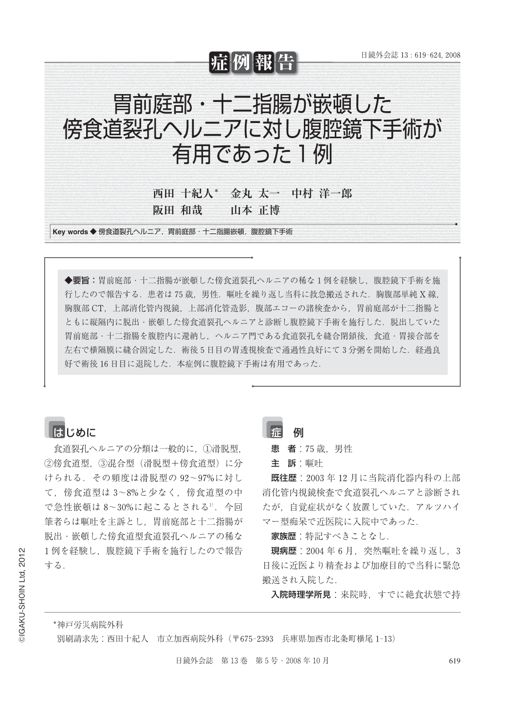
◆要旨:胃前庭部・十二指腸が嵌頓した傍食道裂孔ヘルニアの稀な1例を経験し,腹腔鏡下手術を施行したので報告する.患者は75歳,男性.嘔吐を繰り返し当科に救急搬送された.胸腹部単純X線,胸腹部CT,上部消化管内視鏡,上部消化管造影,腹部エコーの諸検査から,胃前庭部が十二指腸とともに縦隔内に脱出・嵌頓した傍食道裂孔ヘルニアと診断し腹腔鏡下手術を施行した.脱出していた胃前庭部・十二指腸を腹腔内に還納し,ヘルニア門である食道裂孔を縫合閉鎖後,食道・胃接合部を左右で横隔膜に縫合固定した.術後5日目の胃透視検査で通過性良好にて3分粥を開始した.経過良好で術後16日目に退院した.本症例に腹腔鏡下手術は有用であった.
We herein reported a rare case of paraesophageal hiatus hernia with incarceration of the antrum and duodenum treated by laparoscopic surgery. A 75-year-old male was emergently transferred to our hospital for continuous vomiting. The patient was diagnosed as paraesophageal hernia with incarceration of the antrum and duodenum by the examinations of chest-abdominal X-ray, chest-abdominal computed tomography, abdominal ultrasonography, gastric fiberscopy and esophago-gastric image. Gastroesophageal reflux disease(GERD)was not revealed. Laparoscopic surgery was performed by 4 trocar ports. The incarcerating antrum and duodenum were easily pulled back to the abdominal space. The orifice of paraesophagel hiatus hernia was closed by suture, and both sides of the esophago-gastric junction were sutured to the diaphragm. After esophago-gastric image on post-operative day 5, which revealed function of lower esophageal sphincter(LES)and passage from esophagus to duodenum were normal, feeding was started. The patient was discharged on post-operative day 16 without any symptoms. We considered that laparoscopic surgery for paraesophageal hiatus hernia was effective.

Copyright © 2008, JAPAN SOCIETY FOR ENDOSCOPIC SURGERY All rights reserved.


