Japanese
English
- 有料閲覧
- Abstract 文献概要
- 1ページ目 Look Inside
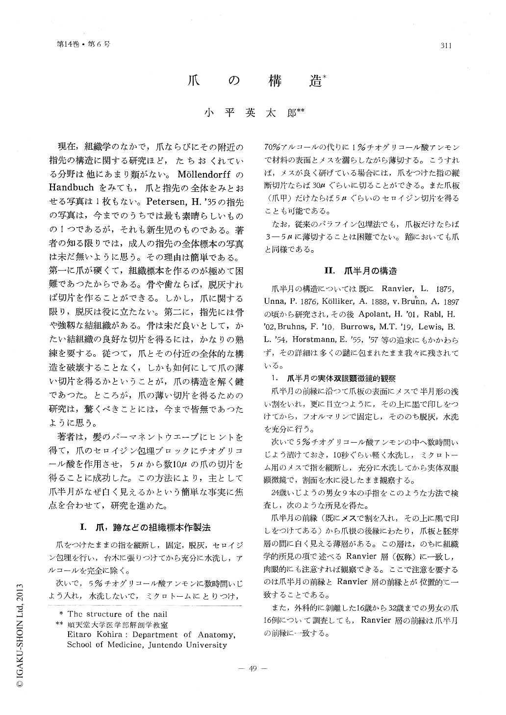
ヒトの手の指を縦断し,その断面を実体双眼顕微鏡で検査した。次いで爪のセロイジン包埋材料をチオグリコール酸アンモンに漬けて,5-数10μの組織標本を作り,また従来の方法で5μ以下のパラフイン切片を作つた。更にまた一部は電子顕微鏡で観察し,次のような所見を得た。
爪半月の前縁から爪根の後縁にわたり,爪板と胚芽層の間に,肉眼的に白く見える薄層(仮にRanvier層と名づける)があり,この層の前縁と爪半月の前縁は位置的に一致する。Ranvier層は成人では厚さ60μ,あるいはそれ以上に達し,ここの細胞はSubstance onychogène(Ranvier, L. 1875)という顆粒状の物質を多量に含み,そのため透過光線では褐色に,落下光線では白く輝いて見える。
Substance onychogèneは厚さ5μ以下のパラフィン切片では線維状に見え,Heidenhainの鉄ヘマトキシリンで強く染まり,またエオジン,酸性フクシン,オランゲG,Pearse法でも良く染まる。
電子顕微鏡的にはSubstance onychogèneは直径0.25μ,長径2.5μぐらいの小円柱で,電子密度が大きく,均質に見え,角化張原線維といわれているものに一致する。
以上の所見から次の結論を得た。
1)爪半月が白く見えるのはSubstance onychogène(Ranvier, L. 1875)の乱反射にもとづく。
2)Substance onychogèneは顆粒でなく,角化張原線維である。
Some human fingers were longitudinally cutted, and the sections were examined through a streoscopic microscope. Then, some celloidin embedded materials of nails were immersed in ammonium thioglycolate to make sections of 5 to 30μ or more. In addition, paraffin sections of 5μ or smaller were also made. Further, some other nails were observed through electron microscope. The following is the resuls of these observations:
From the front edge of the nail lunula extending to the hind edge of the nail root, there is a thin layer (let us temporarily call it Ranvier's layer) which looks white macroscopically. The front edge of this layer falls exactly on the front edge of the nail lunula. The Ranvier's layer of an adult is 60μ or more in thickness, and its cells contain a large quantity of granular substance which is called substance onychogène (Ranvier, L. 1875). This fact makes this layer looks brown by the transmitting light and brilliantly white by the fall light.
Stubstance onychogène in a paraffin section of 5μ or less in thickness presents a delicate fibrous form. It is intensely stained in Heidenhain's iron-hematoxylin. It is also stained well in eosin, acid-fuchsin and orange G. Furthermore, it is stained specifically with Pearse's staining method.
Electronmicroscopically observed, substance onychogène is a small column of about 0.25μ in short diameter and 2.5μ in long diameter. It has a high electron density, and looks homogeneous. It is identified as what is called keratinized tonofibril.
From the above observation the following conclusions are obtained:
1) The whiteness of the nail lunula is due to the irregular reflection of substance onychogène (Ranvier, L. 1875).
2) Substance onchogène is not granules, but keratinized tonofibrils.

Copyright © 1963, THE ICHIRO KANEHARA FOUNDATION. All rights reserved.


