Japanese
English
- 有料閲覧
- Abstract 文献概要
- 1ページ目 Look Inside
- 参考文献 Reference
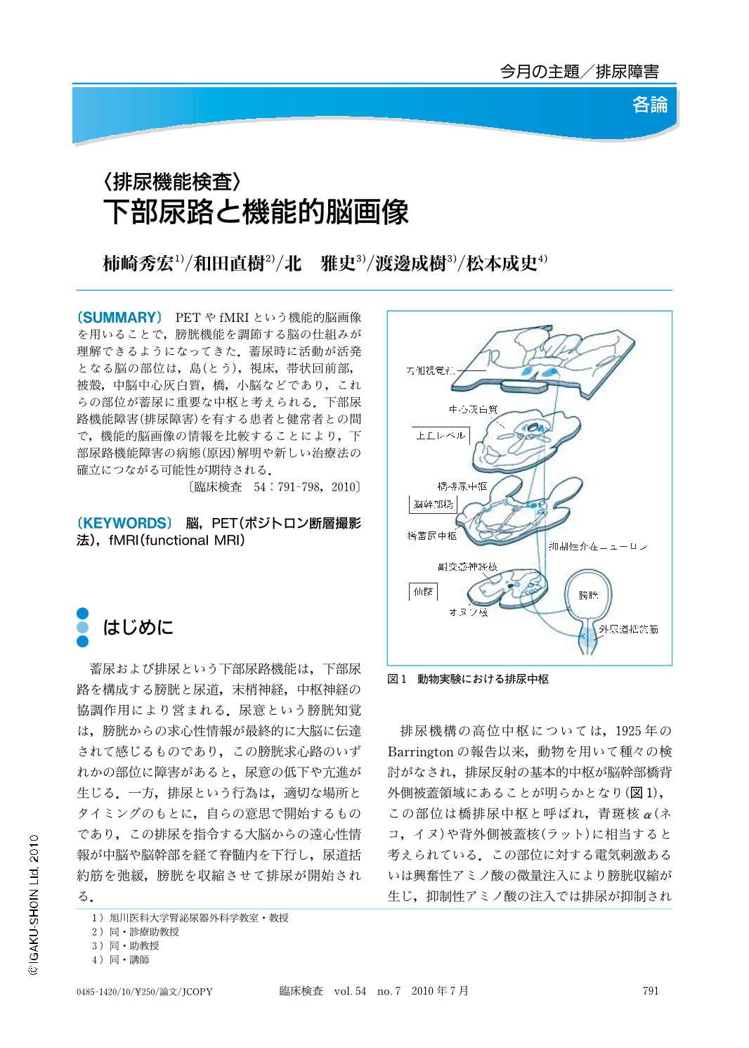
PETやfMRIという機能的脳画像を用いることで,膀胱機能を調節する脳の仕組みが理解できるようになってきた.蓄尿時に活動が活発となる脳の部位は,島(とう),視床,帯状回前部,被殻,中脳中心灰白質,橋,小脳などであり,これらの部位が蓄尿に重要な中枢と考えられる.下部尿路機能障害(排尿障害)を有する患者と健常者との間で,機能的脳画像の情報を比較することにより,下部尿路機能障害の病態(原因)解明や新しい治療法の確立につながる可能性が期待される.
Functional brain imaging such as PET (positron emission tomography) and fMRI (functional MRI) is a very useful tool for examining how the brain controls bladder function in humans. Recent studies with functional brain imaging have shown several brain regions that are relevant to bladder control, including insula, thalamus, anterior cingulate gyrus, putamen, periaqueductal gray, pons and cerebellum. By comparing functional brain imaging between healthy volunteers and patients with lower urinary tract dysfunction, we will be able to promote our understanding of the pathophysiology of lower urinary tract dysfunction and a new therapeutic strategy.

Copyright © 2010, Igaku-Shoin Ltd. All rights reserved.


