Japanese
English
- 有料閲覧
- Abstract 文献概要
- 1ページ目 Look Inside
I.はじめに
頭蓋内dermoid cystは,胎生3-4週の神経管形成時の閉鎖不全に起因する先天性脳腫瘍であり1,5,8,10),その頻度は日本脳腫瘍統計によれば,0.2%と稀な腫瘍である12).さらに,頭蓋内テント上dermoid cystに関しては臨床的検討を加えたいくつかの報告はあるものの,海綿静脈洞部に発生したdermoid cystの報告例は極めて稀である3,7-9).最近,動眼神経麻痺で発症し,海綿静脈洞壁より発生したと考えられた傍トルコ鞍部dermoidcystの1例を経験したので,文献的考察を加えて報告する.
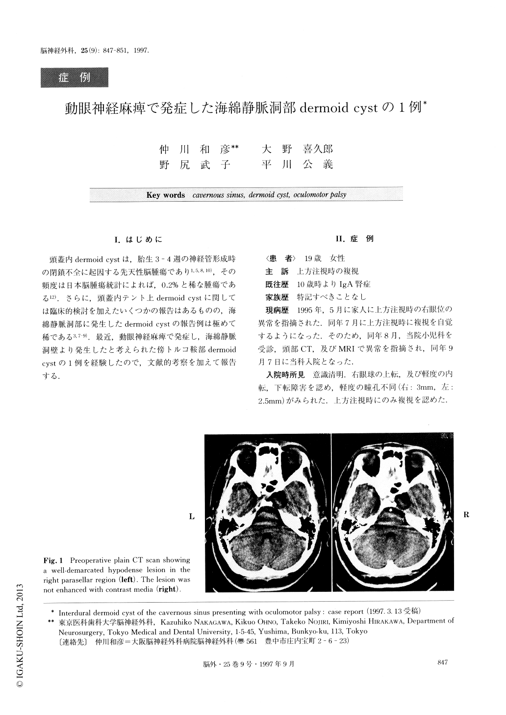
A 19-year-old female was admitted to our hospital because of diplopia. Neurological examination showed slight anisocoria (right>left), disturbance of ocular movement in upward, downward and medial directions on the right side, and diplopia on upward gaze. Com-puted tomography (CT) scan demonstrated a well-demarcated cystic lesion in the right parasellar region. The lesion showed non-homogeneous low intensity with a high intensity margin on Tl-weighted imagingand high intensity with a low intensity margin on T2-weighted imaging in magnetic resonance image (MRI).The lesion showed enhancement by contrast media on neither CT nor MRI. Cerebral angiography revealed no tumor stains. The lesion was approached via right fron-to-temporal craniotomy. The right optic nerve, oculo-motor nerve and internal carotid artery were compress-ed medially by the tumor. The tumor contents appeared to be flaky and greasy containing motor-oil-like fluid, mineralized body, and a few hairs. The tumor was attached to the lateral wall of the right cavernous sinus (inner membranous layer), but was totally resected with careful microscopic dissection.
The histopathological examination confirmed the di-agnosis of dermoid cyst. The patient's complaint of di-plopia showed gradual improvement postoperatively. Supratentorial intracranial dermoid cyst is a rare con-genital tumor and, to our knowledge, only one case arising within the lateral wall of the cavernous sinus and presenting oculomotor palsy has been reported. Total removal is ideal and may be possible even when the tumor arises from the lateral wall of the cavernous sinus.

Copyright © 1997, Igaku-Shoin Ltd. All rights reserved.


