Japanese
English
- 有料閲覧
- Abstract 文献概要
- 1ページ目 Look Inside
I.はじめに
脳動脈瘤に対するクリッピング術に代わって塞栓術で治療しようとする試みは,古くはMullan6)により報告され種々の塞栓物質,塞栓方法が発表されてきている.中でもGuglielmiら3,4)が1991年に発表したdetachablecoilは,血管内腔より使用できる画期的なもので,現在その臨床応用が広く行われつつある.しかしこの治療法は,動物実験では治療後の治癒過程を検討されてはいるが,臨床症例での病理学的検討は十分なされているとは言えない.
われわれの施設では,開頭手術が困難と考えられる高齢者や重症者に限って,detachable coilを用いた脳動脈瘤の塞栓術を行っているが,今回くも膜下出血で発症しcoil塞栓術を施行後,全身合併症で死亡した1例を剖検する機会を得たので,その病理学的所見を報告し,文献的考察を加える.
We described an autopsy case of a ruptured aneurys-mal subarachnoid hemorrhage treated with endovascu-lar embolization by interlocking detachable coils.
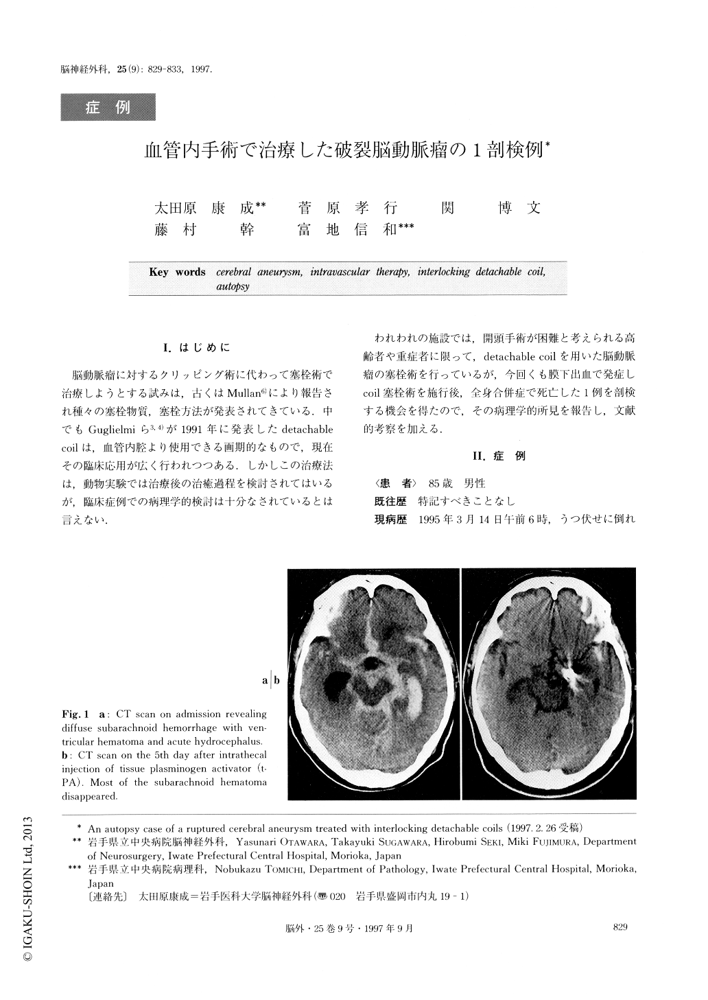
An 85-year-old male presented with sudden onset of severe subarachnoid hemorrhage. Cerebral angiogram revealed a right internal carotid-posterior communicat-ing artery aneurysm. Post-operative angiogram revealed complete obliteration of the aneurysm, except for its orifice. Following the embolization of the aneurysm, tis-sue plasminogen activator was intrathecally perfused for anti-vasospasm treatment. Follow-up angiogram showed stable obliteration of the aneurysm, and no par-ticular findings of cerebral vasospasm. The patient had been recovering without any neurological deficits, but died from pneumonia on the 25th day after the embo-lization.
Autopsy findings revealed the disappearance of thesubarachnoid hemorrhage, and no visible finding of cerebral infarction or edema. The inner lumen of the aneurysm was occupied by a mixture of the coils and the clots. The surface of the embolized coils was direct-ly observed through the orifice of the aneurysm with-out any membranous substance from the inner lumen of the internal carotid artery.
This pathological finding is different from the pre-viously reported animal models in which the surface of the embolized coils was covered with endothelial mem-brane 2 weeks after embolization. Further examinations are required to clarify the pathogenesis of the endothe-lial regrowth.

Copyright © 1997, Igaku-Shoin Ltd. All rights reserved.


