Japanese
English
- 有料閲覧
- Abstract 文献概要
- 1ページ目 Look Inside
I.はじめに
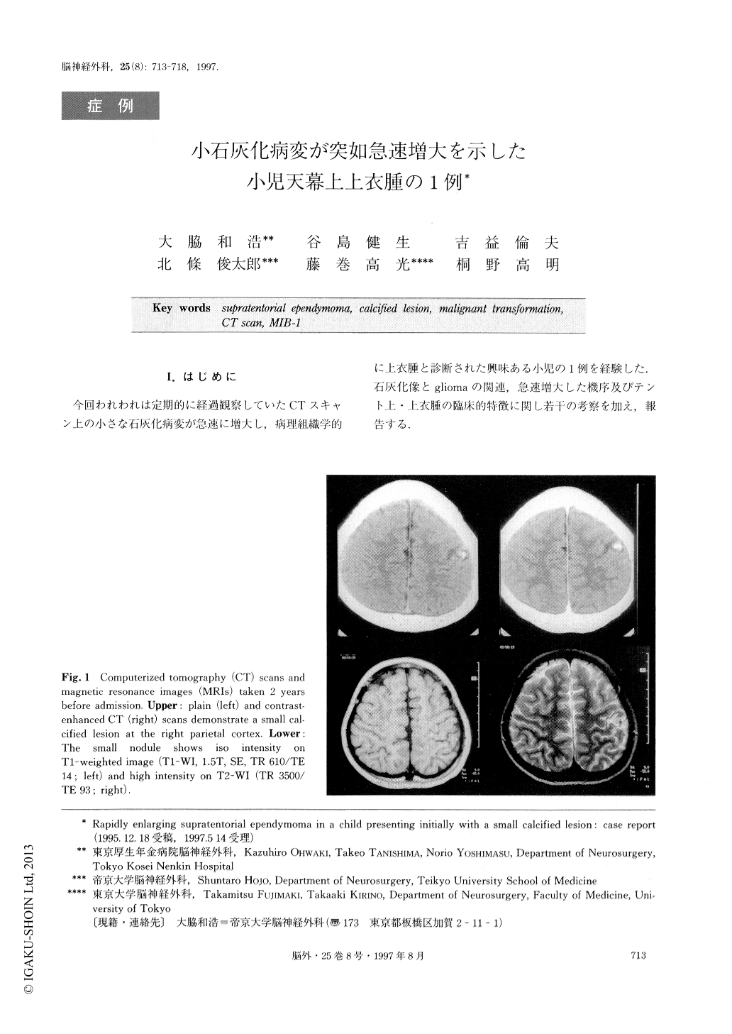
今回われわれは定期的に経過観察していたCTスキャン上の小さな石灰化病変が急速に増大し,病理組織学的に上衣腫と診断された興味ある小児の1例を経験した.石灰化像とgliomaの関連,急速増大した機序及びテント上・上衣腫の臨床的特徴に関し若干の考察を加え,報告する.
The authors report an unusual case of a 11-year-old boy whose supratentorial ependymoma showed rapid growth. He had had generalized convulsive seizures when he was 9 years old. On an initial CT scan a small calcified lesion was identified adjacent to the right sen-sorimotor cortex. Repeated CT scans showed no inter-val change in the size of the tumor for 16 months. Then, he suffered an acute onset of left hemiparesis. The neuroimaging studies demonstrated a huge tumor with a large cyst in the right parietal region which had not been observed on CT scan 7 months before. Total removal of the tumor was performed and the histo-pathological diagnosis was ependymoma with no evi-dence of malignancy. However, MIB-1 staining of the specimen revealed a high index of proliferative poten-tial up to 25% in some area. The high score of MIB-1 staining correlated well with the rapid clinical course of this histologically benign ependymoma. The small calci-fied lesion demonstrated on the initial CT scan in this case is considered to have been a low grade ependymo-ma and to have abruptly transformed into a higher grade, one resulting in rapid enlargement. The authors stress that small intracranial calcified lesions should be carefully followed up by repeated neuroimaging studies at short intervals.

Copyright © 1997, Igaku-Shoin Ltd. All rights reserved.


