Japanese
English
- 有料閲覧
- Abstract 文献概要
- 1ページ目 Look Inside
I.はじめに
くも膜下出血の中には血管撮影を施行しても出血源を明らかにできない症例がある.これらの一部は脳血管撮影を繰り返すことにより動脈瘤が発見されることもあるが多くは原因不明のままである3,6,9,12,13).
今回,われわれは初回血管撮影で動脈瘤の虚脱のため血管撮影で造影されず,さらに破裂部に形成された嚢状動脈瘤様のblebが2回目の血管撮影で確認され手術に至った破裂内頸—後交通動脈瘤の症例を経験したので若干の考察を含め報告する.
A case of saccular aneurysm-like bleb formation after rupture of an aneurysm in the internal carotid-posterior communicating artery. In connection with this, an in-teresting angiographic finding was reported.
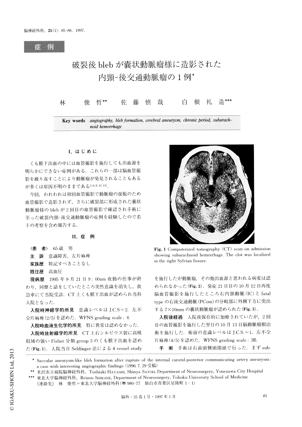
A 65-year-old man suffered from sudden disturbance of consciousness and left hemiparesis. Computed tomography (CT) scan revealed typical subarachnoid hemorrhage localized in the right Sylvian fissure. Next day, a cerebral angiography was performed, but no aneurysm was detected. A second angiography was performed 21 days after the onset, and it revealed a saccular right internal carotid-posterior communicating artery (IC-PC) aneurysm. An operation for the IC-PC aneurysm was performed by conventional pterional approach. However, intraoperative findings were unex-pected. The collapsed ruptured true IC-PC aneurysm was found at the orifice of the larger bleb, and the rup-tured point was in the neck of the true aneurysm. Clip-ping of the aneurysm was performed successfully. The patient was discharged on foot. The aneurysm detected by the second angiography was not a true one but a bleb formation. Continuous hemodynamic stress on the rupture point may induce the formation of such an aneurysm-like bleb.
It should be kept in mind that an aneurysm found in the chronic period might be an aneurysm-like bleb.

Copyright © 1997, Igaku-Shoin Ltd. All rights reserved.


