Japanese
English
- 有料閲覧
- Abstract 文献概要
- 1ページ目 Look Inside
I.はじめに
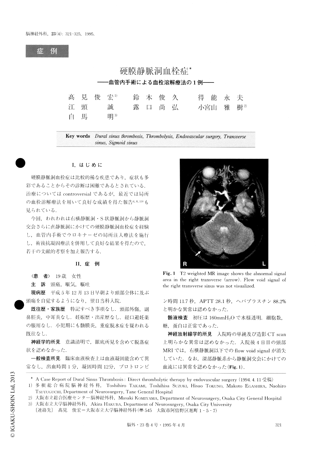
硬膜静脈洞血栓症は比較的稀な疾患であり,症状も多彩であることからその診断は困難であるとされている.治療についてはcontroversialであるが,最近では局所の血栓溶解療法を用いて良好な成績を得た報告4,8,10)も見られている.
今回,われわれは右横静脈洞・S状静脈洞から静脈洞交会さらに直静脈洞にかけての硬膜静脈洞血栓症を経験し,血管内手術でウロキナーゼの局所注入療法を施行し,術後抗凝固療法を併用して良好な結果を得たので,若干の文献的考察を加え報告する.
Dural sinus thrombosis, a relatively rare disease, is difficult to diagnose because of variable symptomatic manifestations. We successfully treated a case of dural sinus thrombosis by direct thrombolysis using an en-dovascular technique in combination with postoperative anticoagulant therapy.
The patient, a 19-year-old female, developed a headache affecting her whole head on December 13, 1993. She was admitted to our hospital the next day. Neurological examination upon admission revealed no neurological abnormalities, nor was there any abnor-mality in CT scan, either plain or enhanced, taken on the day of admission. The patient's consciousness de-teriorated in the early morning of December 22. MRI and cerebral angiography revealed thrombi from the confluence of the sinuses to the right transverse and sigmoid sinus, with disturbed circulation through deep cerebral veins. Systemic thrombolytics, steroid and mannitol were started, but, on the next day, the third ventricle was compressed by bilateral swelling of the basal ganglia, with hydrocephalus. Since her conscious-ness deteriorated further despite ventricular drainage and barbiturate therapy, direct thrombolytic therapy was performed on December 25. A catheter was placed in the superior sagittal sinus, and 600,000 units of uro-kinase was locally injected, followed by postoperative anticoagulant therapy. The patient's condition improved rapidly. On CT scan, the bilateral swelling of the basal ganglia disappeared along with the hydrocephalus. At about 1 month after endovascular surgery, MRI and cerebral angiography revealed recanalization of the deep cerebral veins, straight sinus and confluence of sinuses with improved opacification of the left trans-verse sinus, although the right transverse sinus was found to be re-occluded. On February 20, the patient was discharged without any neurological deficits.
Copyright © 1995, Igaku-Shoin Ltd. All rights reserved.