Japanese
English
- 有料閲覧
- Abstract 文献概要
- 1ページ目 Look Inside
- 参考文献 Reference
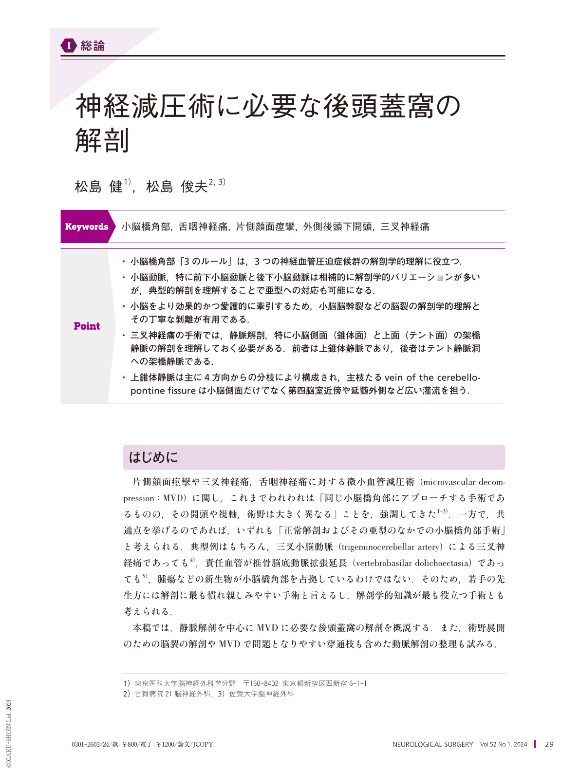
Point
・小脳橋角部「3のルール」は,3つの神経血管圧迫症候群の解剖学的理解に役立つ.
・小脳動脈,特に前下小脳動脈と後下小脳動脈は相補的に解剖学的バリエーションが多いが,典型的解剖を理解することで亜型への対応も可能になる.
・小脳をより効果的かつ愛護的に牽引するため,小脳脳幹裂などの脳裂の解剖学的理解とその丁寧な剝離が有用である.
・三叉神経痛の手術では,静脈解剖,特に小脳側面(錐体面)と上面(テント面)の架橋静脈の解剖を理解しておく必要がある.前者は上錐体静脈であり,後者はテント静脈洞への架橋静脈である.
・上錐体静脈は主に4方向からの分枝により構成され,主枝たるvein of the cerebellopontine fissureは小脳側面だけでなく第四脳室近傍や延髄外側など広い灌流を担う.
In most microvascular decompression surgeries, surgical maneuvers are performed within normal anatomical structures without any neoplasms. Thus, detailed anatomical knowledge is essential to perform safe and efficient procedures. “Rule of 3” by Rhoton AL Jr. is helpful for understanding not only the anatomy of the posterior fossa but also the three neurovascular compression syndromes. The cerebellar arteries and posterior fossa veins have substantial variability, but a basic understanding of their typical patterns is useful to explore individual cases. To use adequate surgical approaches through the cerebellar tentorial or petrosal surface in individual trigeminal neuralgia surgeries, anatomical knowledge of the bridging veins on the tentorial(the bridging veins into the tentorial sinus)and petrosal surfaces(the superior petrosal vein)is crucial. Fissure openings help to minimize cerebellar retraction, similarly to the sylvian fissure dissection in supratentorial surgeries.

Copyright © 2024, Igaku-Shoin Ltd. All rights reserved.


