Japanese
English
- 有料閲覧
- Abstract 文献概要
- 1ページ目 Look Inside
- 参考文献 Reference
Ⅰ.はじめに
脳神経外科手術後に予想外の視力障害を来した報告は散見されるが,正確な病態が明らかではない症例も多い5).今回われわれは,未破裂脳動脈瘤開頭術中の偶発的眼窩内進入に起因して眼球後部の液体貯留と眼窩・眼瞼気腫を来し,眼瞼腫脹を伴って視力障害・眼球運動障害等を合併した症例を経験した.後方視的には眼球突出も認められ,眼窩内圧上昇によって脳神経・眼筋等の虚血を来すorbital compartment syndrome(OCS)9,10)の可能性が考えられた.本例の臨床経過を提示し,その病態について考察する.
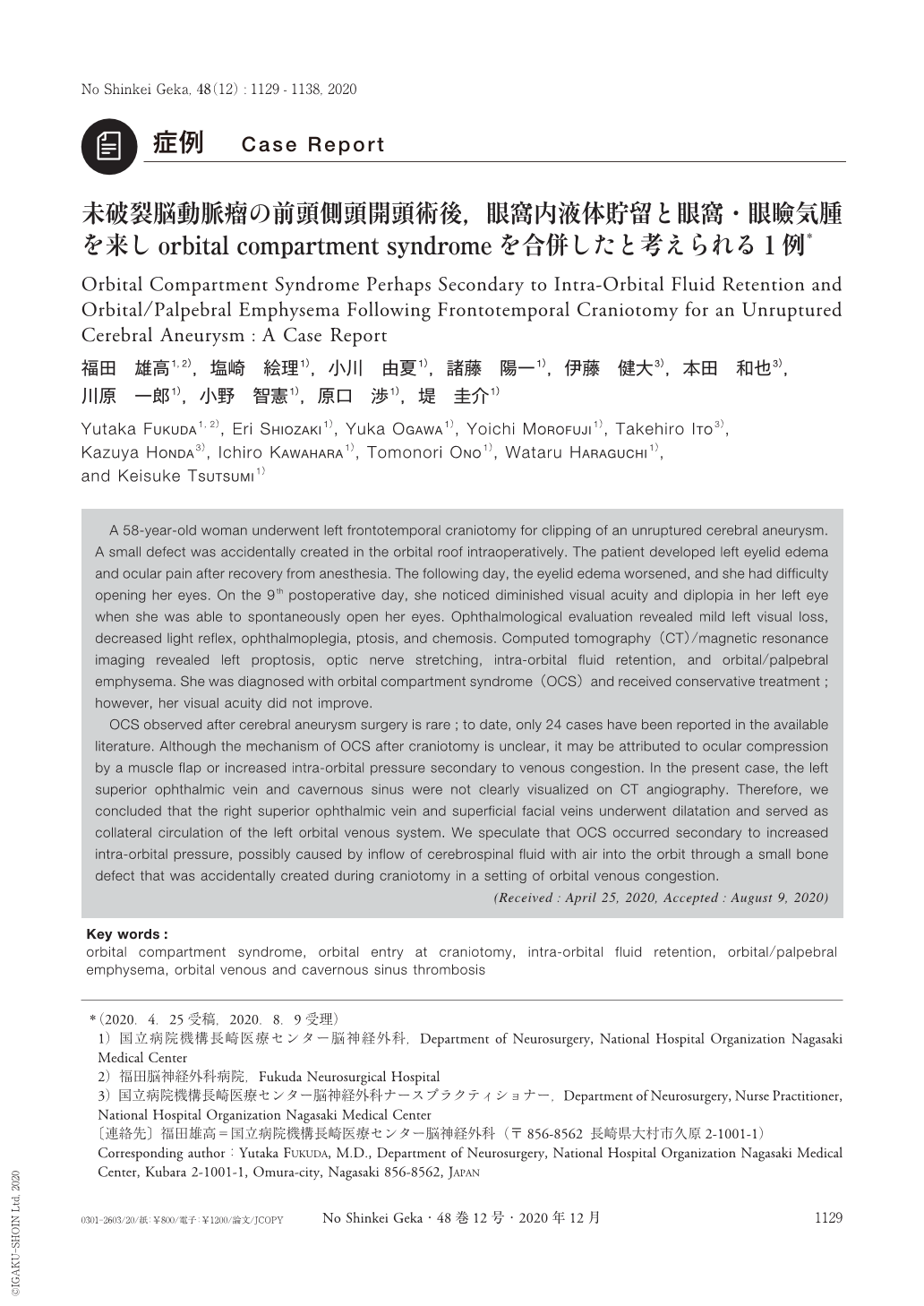
A 58-year-old woman underwent left frontotemporal craniotomy for clipping of an unruptured cerebral aneurysm. A small defect was accidentally created in the orbital roof intraoperatively. The patient developed left eyelid edema and ocular pain after recovery from anesthesia. The following day, the eyelid edema worsened, and she had difficulty opening her eyes. On the 9th postoperative day, she noticed diminished visual acuity and diplopia in her left eye when she was able to spontaneously open her eyes. Ophthalmological evaluation revealed mild left visual loss, decreased light reflex, ophthalmoplegia, ptosis, and chemosis. Computed tomography(CT)/magnetic resonance imaging revealed left proptosis, optic nerve stretching, intra-orbital fluid retention, and orbital/palpebral emphysema. She was diagnosed with orbital compartment syndrome(OCS)and received conservative treatment;however, her visual acuity did not improve.
OCS observed after cerebral aneurysm surgery is rare;to date, only 24 cases have been reported in the available literature. Although the mechanism of OCS after craniotomy is unclear, it may be attributed to ocular compression by a muscle flap or increased intra-orbital pressure secondary to venous congestion. In the present case, the left superior ophthalmic vein and cavernous sinus were not clearly visualized on CT angiography. Therefore, we concluded that the right superior ophthalmic vein and superficial facial veins underwent dilatation and served as collateral circulation of the left orbital venous system. We speculate that OCS occurred secondary to increased intra-orbital pressure, possibly caused by inflow of cerebrospinal fluid with air into the orbit through a small bone defect that was accidentally created during craniotomy in a setting of orbital venous congestion.

Copyright © 2020, Igaku-Shoin Ltd. All rights reserved.


