Japanese
English
- 有料閲覧
- Abstract 文献概要
- 1ページ目 Look Inside
I.はじめに
脊髄髄膜腫の約80%が硬膜内髄外に発生し,残り大部分は硬膜内外にわたって存在する.ところが極めて稀に硬膜外に発生することがあり,硬膜外腫瘍のほとんどが悪性のものであることから,たいていの場合,悪性腫瘍と考えて手術され,術中迅速組織検査や,経験深い術者の洞察によって診断ないし疑いを持たれて根治的切除がなされることが多い.また,術後の固定組織標本によって診断がつき,根治手術が追加される例もある2,6,16).
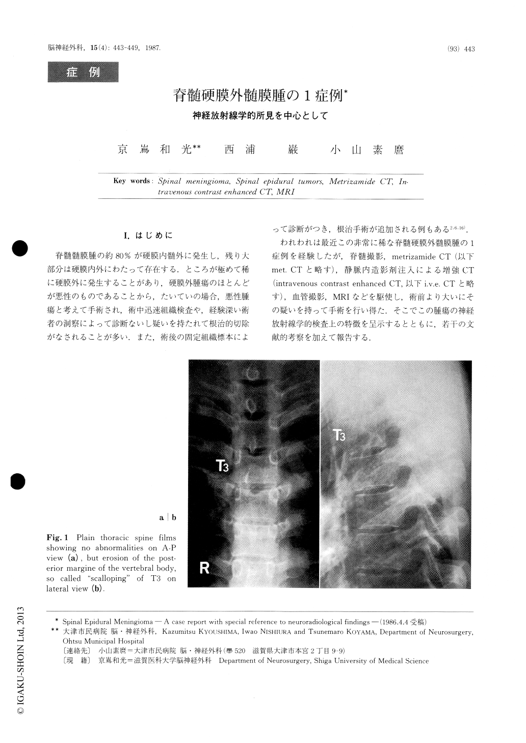
われわれは最近この非常に稀な脊髄硬膜外髄膜腫の1症例を経験したが,脊髄撮影,metrizamide CT (以下met.CTと略す),静脈内造影剤注入による増強CT(intravenous contrast enhanced CT,以下i.v.e.CTと略す),血管撮影,MRIなどを駆使し,術前より大いにその疑いを持って手術を行い得た.そこでこの腫瘍の神経放射線学的検査上の特徴を呈示するとともに,若干の文献的考察を加えて報告する.
Spinal meningiomas are common as intradural-extramedullary neoplasm, but solitaly epidural spinal meningiomas are extremely rare. They may often be misdiagnosed as malignant neoplasms which are much more common in this location. Furthermore, at the time of operation, it is often difficult to distinguish the epidural meningioma from malignant tumors, even by the microscopic examination of the fresh frozen section. We present a case of spinal epidural meningioma, and emphasize the importance of preoperative neuroradiolo-gical examinations.

Copyright © 1987, Igaku-Shoin Ltd. All rights reserved.


