Japanese
English
- 有料閲覧
- Abstract 文献概要
- 1ページ目 Look Inside
- 参考文献 Reference
Ⅰ.はじめに
近年MRIの普及により,日常診療において,頭痛や頚部痛のみを主訴とした椎骨動脈解離に遭遇する機会が増えている.その大部分は予後良好と考えられるが6),稀にくも膜下出血や血管閉塞に移行する症例も報告されている1,4).頭痛・頚部痛以外に神経症状のない患者が,突然重篤な状況に陥る可能性がある以上,その自然歴を明確にして,実際の治療方針に生かすことは重要と考える.これまで当院で経験した症例を後視的に検討し,その臨床的特徴について報告する.
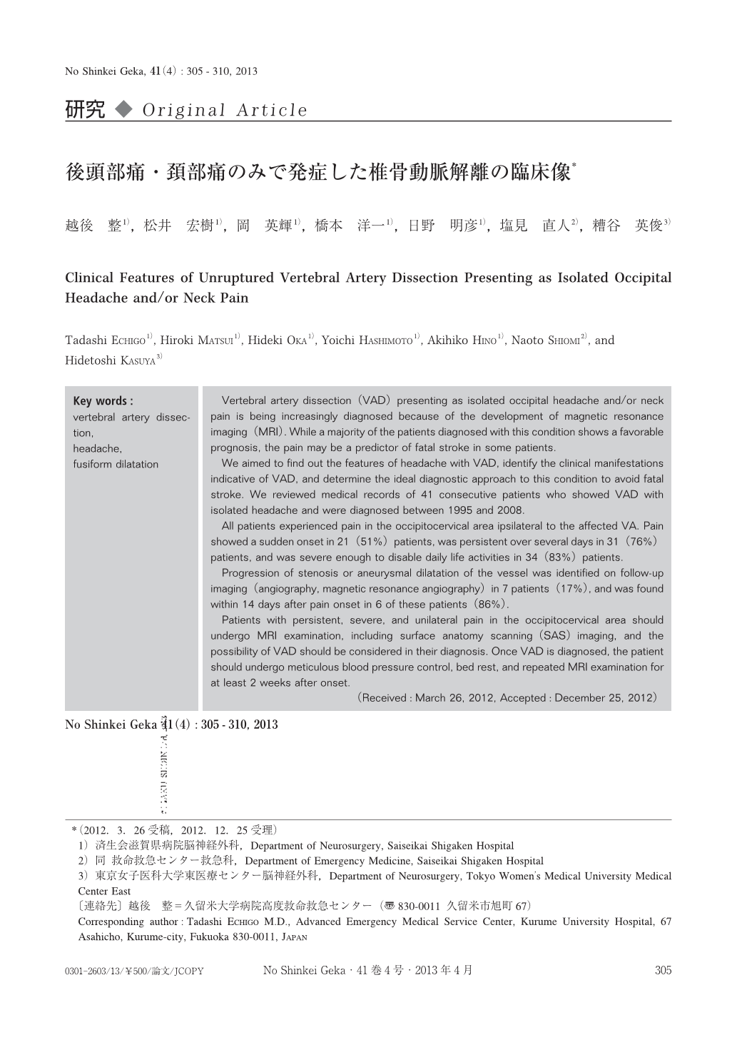
Vertebral artery dissection(VAD)presenting as isolated occipital headache and/or neck pain is being increasingly diagnosed because of the development of magnetic resonance imaging(MRI). While a majority of the patients diagnosed with this condition shows a favorable prognosis, the pain may be a predictor of fatal stroke in some patients.
We aimed to find out the features of headache with VAD, identify the clinical manifestations indicative of VAD, and determine the ideal diagnostic approach to this condition to avoid fatal stroke. We reviewed medical records of 41 consecutive patients who showed VAD with isolated headache and were diagnosed between 1995 and 2008.
All patients experienced pain in the occipitocervical area ipsilateral to the affected VA. Pain showed a sudden onset in 21(51%)patients, was persistent over several days in 31(76%)patients, and was severe enough to disable daily life activities in 34(83%)patients.
Progression of stenosis or aneurysmal dilatation of the vessel was identified on follow-up imaging(angiography, magnetic resonance angiography)in 7 patients(17%), and was found within 14 days after pain onset in 6 of these patients(86%).
Patients with persistent, severe, and unilateral pain in the occipitocervical area should undergo MRI examination, including surface anatomy scanning(SAS)imaging, and the possibility of VAD should be considered in their diagnosis. Once VAD is diagnosed, the patient should undergo meticulous blood pressure control, bed rest, and repeated MRI examination for at least 2 weeks after onset.

Copyright © 2013, Igaku-Shoin Ltd. All rights reserved.


