Japanese
English
- 有料閲覧
- Abstract 文献概要
- 1ページ目 Look Inside
- 参考文献 Reference
Ⅰ.はじめに
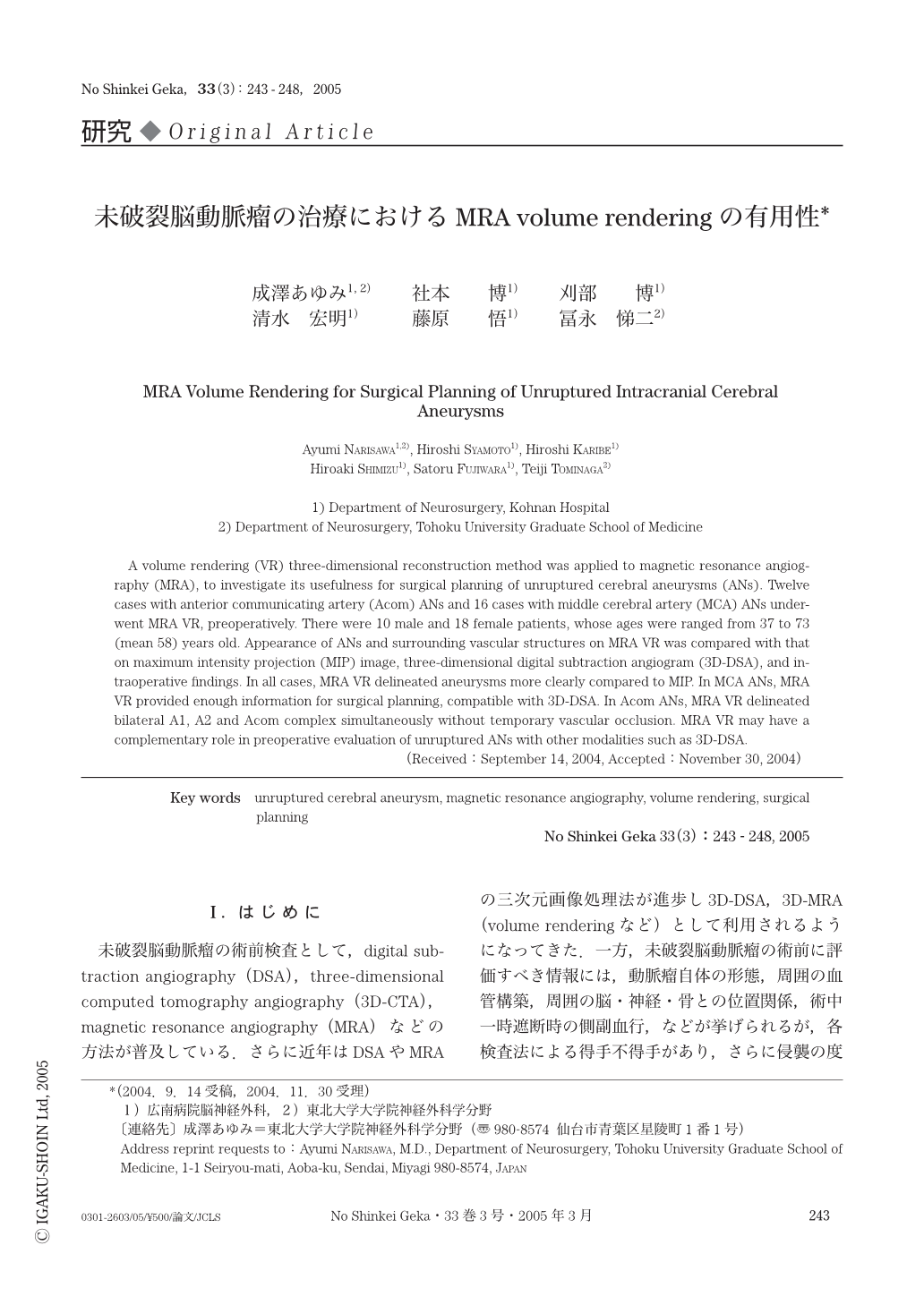
未破裂脳動脈瘤の術前検査として,digital subtraction angiography(DSA),three-dimensional computed tomography angiography(3D-CTA),magnetic resonance angiography(MRA)などの方法が普及している.さらに近年はDSAやMRAの三次元画像処理法が進歩し3D-DSA,3D-MRA(volume renderingなど)として利用されるようになってきた.一方,未破裂脳動脈瘤の術前に評価すべき情報には,動脈瘤自体の形態,周囲の血管構築,周囲の脳・神経・骨との位置関係,術中一時遮断時の側副血行,などが挙げられるが,各検査法による得手不得手があり,さらに侵襲の度合いも異なる.特に,最近臨床応用可能となったMRA volume rendering(以下MRA VR)については,その有用性や限界に関する報告は少ない.そこで今回,未破裂脳動脈瘤における術前検査としての有用性を検討したので報告する.
A volume rendering (VR) three-dimensional reconstruction method was applied to magnetic resonance angiography (MRA), to investigate its usefulness for surgical planning of unruptured cerebral aneurysms (ANs). Twelve cases with anterior communicating artery (Acom) ANs and 16 cases with middle cerebral artery (MCA) ANs underwent MRA VR, preoperatively. There were 10 male and 18 female patients, whose ages were ranged from 37 to 73 (mean 58) years old. Appearance of ANs and surrounding vascular structures on MRA VR was compared with that on maximum intensity projection (MIP) image, three-dimensional digital subtraction angiogram (3D-DSA), and intraoperative findings. In all cases,MRA VR delineated aneurysms more clearly compared to MIP. In MCA ANs, MRA VR provided enough information for surgical planning, compatible with 3D-DSA. In Acom ANs, MRA VR delineated bilateral A1, A2 and Acom complex simultaneously without temporary vascular occlusion. MRA VR may have a complementary role in preoperative evaluation of unruptured ANs with other modalities such as 3D-DSA.

Copyright © 2005, Igaku-Shoin Ltd. All rights reserved.


