Japanese
English
- 有料閲覧
- Abstract 文献概要
- 1ページ目 Look Inside
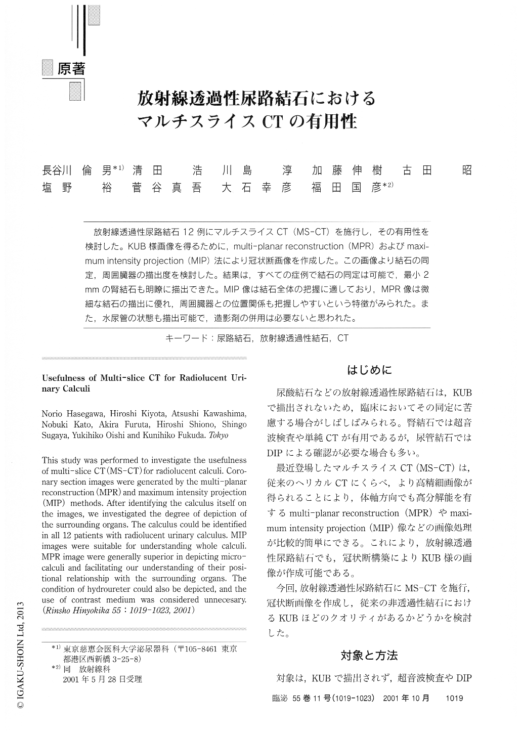
放射線透過性尿路結石12例にマルチスライスCT(MS-CT)を施行し,その有用性を検討した。KUB様画像を得るために,multi-planar reconstruction(MPR)およびmaxi-mum intensity projection(MIP)法により冠状断画像を作成した。この画像より結石の同定,周囲臓器の描出度を検討した。結果は,すべての症例で結石の同定は可能で,最小2mmの腎結石も明瞭に描出できた。MIP像は結石全体の把握に適しており,MPR像は微細な結石の描出に優れ,周囲臓器との位置関係も把握しやすいという特徴がみられた。また,水尿管の状態も描出可能で,造影剤の併用は必要ないと思われた。
This study was performed to investigate the usefulnessof multi-slice CT (MS-CT) for radiolucent calculi. Coro-nary section images were generated by the multi-planarreconstruction (MPR) and maximum intensity projection(MIP) methods. After identifying the calculus itself onthe images, we investigated the degree of depiction ofthe surrounding organs. The calculus could be identifiedin all 12 patients with radiolucent urinary calculus. MIPimages were suitable for understanding whole calculi.

Copyright © 2001, Igaku-Shoin Ltd. All rights reserved.


