Japanese
English
- 有料閲覧
- Abstract 文献概要
- 1ページ目 Look Inside
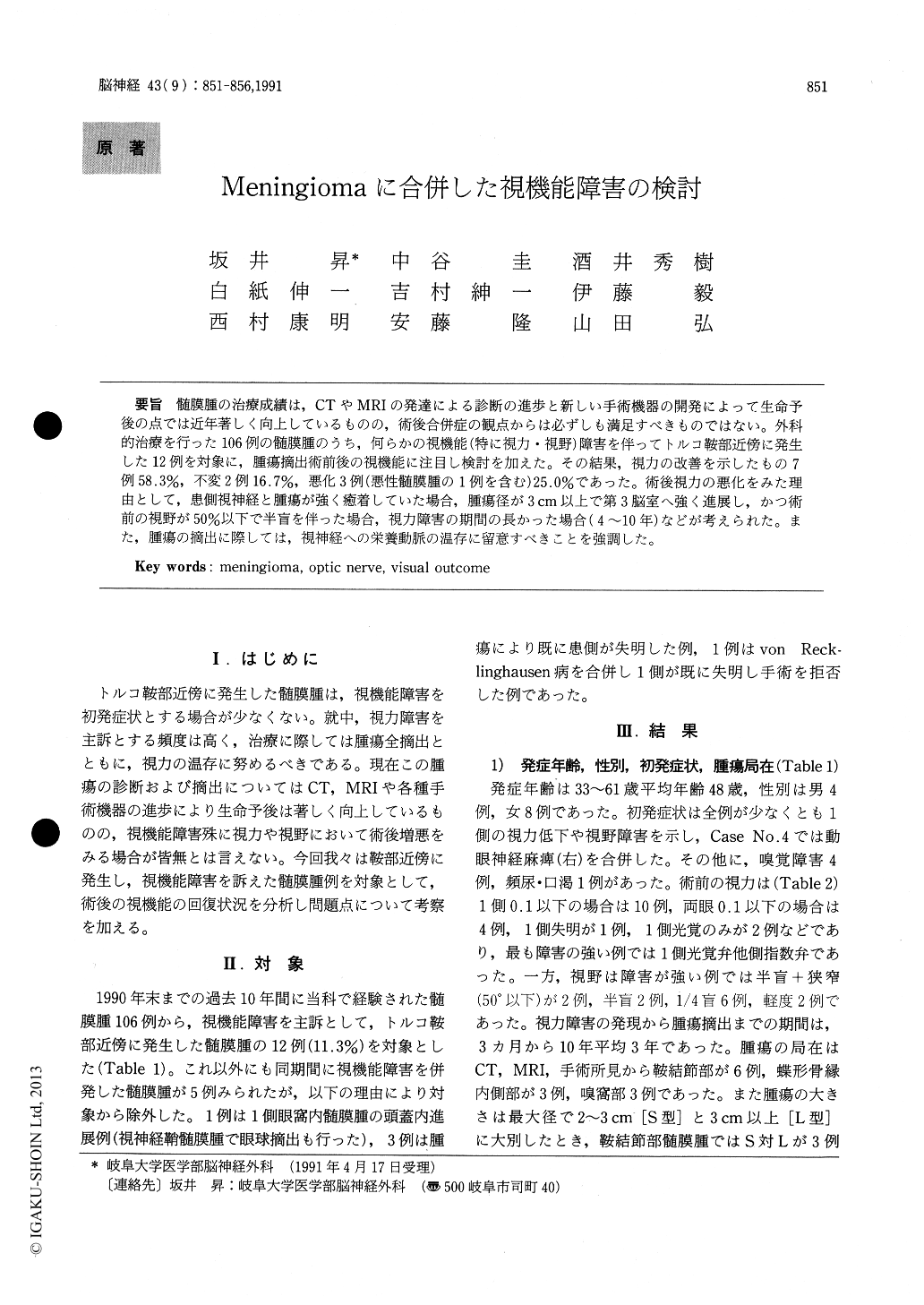
髄膜腫の治療成績は,CTやMRIの発達による診断の進歩と新しい手術機器の開発によって生命予後の点では近年著しく向上しているものの,術後合併症の観点からは必ずしも満足すべきものではない。外科的治療を行った106例の髄膜腫のうち,何らかの視機能(特に視力・視野)障害を伴ってトルコ鞍部近傍に発生した12例を対象に,腫瘍摘出術前後の視機能に注目し検討を加えた。その結果,視力の改善を示したもの7例58.3%,不変2例16.7%,悪化3例(悪性髄膜腫の1例を含む)25.0%であった。術後視力の悪化をみた理由として,患側視神経と腫瘍が強く癒着していた場合,腫瘍径が3cm以上で第3脳室へ強く進展し,かつ術前の視野が50%以下で半盲を伴った場合,視力障害の期間の長かった場合(4〜10年)などが考えられた。また,腫瘍の摘出に際しては,視神経への栄養動脈の温存に留意すべきことを強調した。
Among 106 patients of meningioma surgically experienced the past 10 years between 1981 and 1990, twelve of meningioma with progressive visual impairment were analyzed in relation to postoper-ative visual outcome. There were four males and eight females, and the age ranged from 33 to 61 years with the average 48 years. The distribution of tumor location was 6 cases in tuberculum sellae, 3 cases in the inner side of sphenoid ridge, and 3 cases in olfactory groove. The size of tumor in each case was 2 to 7cm in diameter, and in 8 cases more than 3cm. The duration of visual disturbance was between 3 months and 10 years with the average 3 years. For all cases, surgical removal of the tumor was performed totally by pterional and bifrontal approach. Consequently, 58.3% of 7 cases had im-proved vision postoperatively, 16.7% of 2 cases remained unchanged, and 25.0% of 3 cases were worse, including one case of malignant meningioma, Visual outcome was mainly affected by a duration of symptoms, a tumor size, a preopertive visual impairment, and in special, a situation of optic nerve where compression of tumor itself and adher-ence to the surrounding tissues took place. On oper-ation, great care should be paid for a case of long-standing, severe visual disturbance as demonstrat-ing hemianopsia with visual narrowing less than 50 degree by perimetry, and also for preservation of the feeding arteries of optic nerves.

Copyright © 1991, Igaku-Shoin Ltd. All rights reserved.


