Japanese
English
- 有料閲覧
- Abstract 文献概要
- 1ページ目 Look Inside
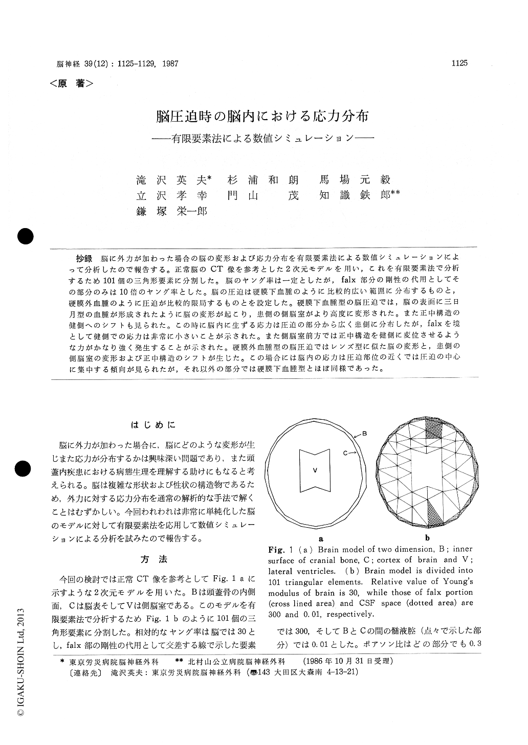
抄録 脳に外力が加わった場合の脳の変形および応力分布を有限要素法による数値シミュレーションによって分析したので報告する。正常脳のCT像を参考とした2次元モデルを用い,これを有限要素法で分析するため101個の三角形要素に分割した。脳のヤング率は一定としたが,falx部分の剛性の代用としてその部分のみは10倍のヤング率とした。脳の圧迫は硬膜下血腫のように比較的広い範囲に分布するものと,硬膜外血腫のように圧迫が比較的限局するものとを設定した。硬膜下血腫型の脳圧迫では,脳の表面に三日月型の血腫が形成されたように脳の変形が起こり,患側の側脳室がより高度に変形された。また正中構造の健側へのシフトも見られた。この時に脳内に生ずる応力は圧迫の部分から広く患側に分布したが,falxを境として健側での応力は非常に小さいことが示された。また側脳室前方では正中構造を健側に変位させるような力がかなり強く発生することが示された。硬膜外血腫型の脳圧迫ではレンズ型に似た脳の変形と,患側の側脳室の変形および正中構造のシフトが生じた。この場合には脳内の応力は圧迫部位の近くでは圧迫の中心に集中する傾向が見られたが,それ以外の部分では硬膜下血腫型とほぼ同様であった。
The analysis of the deformation of brain and distribution of stress caused by the compression from outside would be interesting and also useful for the better understanding of the pathophysio-logy in the cases of intracranial hematoma and other space occupying lesions. The computerized numerical simulation using the finite element me-thod was carried out to analyze these problems. A simple brain model of two dimensions, that was composed of the inner surface of skull, brain and lateral ventricles, was utilized and it was divided into 101 triangular elements for the calculation of finite element method by a 16-bit personal compu-ter. The model of brain was compressed in two different ways ; one was a subdural hematoma type where the force of compression was distributed over relatively wide area and the other was a epidural hematoma type where compression was localized to smaller area. In both types, the brain was compressed on the left fronto-temporal region.
The deformation of brain in the subdural hema-toma type model was that corresponding to the hematoma of crescent shape just as seen on the CT scan of actual case. The lateral ventricle of affect-ed side was deformed more markedly than that of contralateral side. The midline structure of the brain was shifted to normal side and shift was larger in the portion anterior to the lateral ven-tricles. The stress was distributed from the area beneath the hematoma to remote location in the affected hemisphare but the propergation of stress was blocked by the falx and it was very small in the contralateral hemisphare. The concentration of stress was noted in the area anterior to the lateral ventricles and it was seem to correspond to the more marked shift of the midline structure in that location.
In the epidural hematoma type model, the defor-mation of brain was similar to the case where ahematoma of lens shape was formed over the brain. The lateral ventricle of affected side was deformed more markedly than that of opposite side. The midline structure of the brain was sifted similarly to that of the subdural hematoma type model. The stress distribution in the brain was similar to that in the subdural hematoma type model except for the tendency that the stress was more concent-rated in the area just beneath the compression. These models of brain compression revealed the deformation of brain with characteristics similar to the CT findings of actual cases ; that was consi-dered to verify the validity of the method of com-puter simulation.

Copyright © 1987, Igaku-Shoin Ltd. All rights reserved.


