Japanese
English
- 有料閲覧
- Abstract 文献概要
- 1ページ目 Look Inside
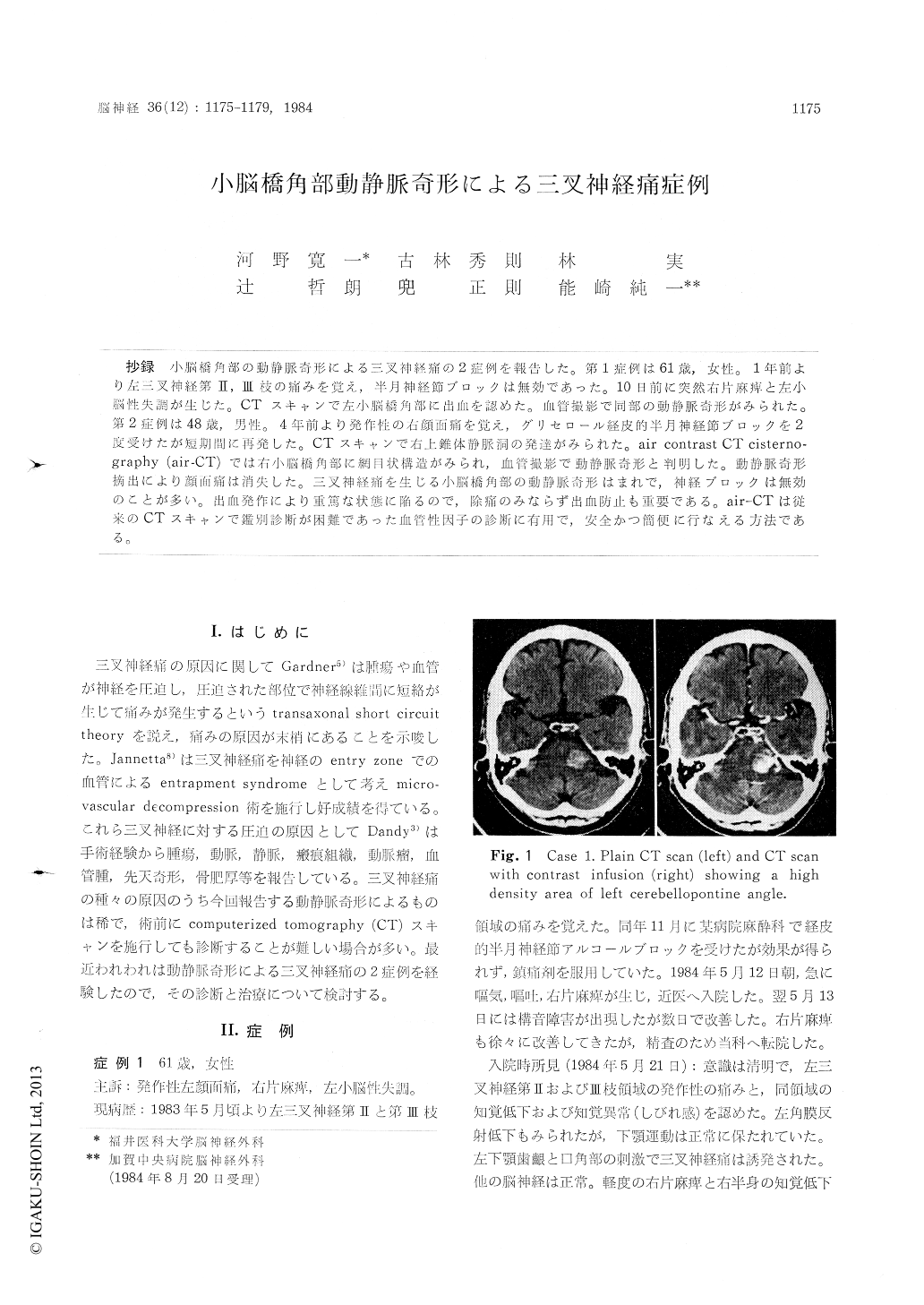
抄録 小脳橋角部の動静脈奇形による三叉神経痛の2症例を報告した。第1症例は61歳,女性。1年前より左三叉神経第II,III枝の痛みを覚え,半月神経節ブロックは無効であった。10日前に突然右片麻痺と左小脳性失調が生じた。CTスキャンで左小脳橋角部に出血を認めた。血管撮影で同部の動静脈奇形がみられた。第2症例は48歳,男性。4年前より発作性の右顔面痛を覚え,グリセロール経皮的半月神経節ブロックを2度受けたが短期間に再発した。CTスキャンで右上錐体静脈洞の発達がみられた。air contrast CT cisterno—graphy (air-CT)では右小脳橋角部に網目状構造がみられ,血管撮影で動静脈奇形と判明した。動静脈奇形摘出により顔面痛は消失した。三叉神経痛を生じる小脳橋角部の動静脈奇形はまれで,神経ブロックは無効のことが多い。出血発作により重篤な状態に陥るので,除痛のみならず出血防止も重要である。air-CTは従来のCTスキャンで鑑別診断が困難であった血管性因子の診断に有用で,安全かつ簡便に行なえる方法である。
Two cases of trigeminal neuralgia caused by arteriovenous malformation (AVM) located in cere-bellopontine angle (CP angle) are presented.
A 61-year-old woman has been suffered from the left 2nd and 3rd division trigeminal neuralgia, and gasserian ganglion rhizotomy did not relieve the pain. Ten days before admission the patient suddenly got right hemiparesis and left cerebellar ataxia. Bleeding of the left CP angle was noticed by computerized tomography (CT) scan and angio-gram revealed AVM of the left CP angle.
A 48-year-old man had right facial neuralgia for 4 years along and he received percutaneous retrogasserian glycerol rhizotomy twice, but the pain was not aleviated. Superior petrosal sinus was enhanced on conventional CT scan, and air cont-rast CT cisternography disclosed network shaped structure at the CP angle, which was revealed as AVM by vertebral angiography. The patient was completely relieved from the neuralgia after re-moval of the AVM.
AVM of the CP angle that causes the trigeminal neuralgia is rare and the gasserian ganglion rhi-zotomy is little effective in aleviating the pain. Bleeding from AVM causes severe neurological deficits. Removal of AVM is important not only for pain relief but also for protecting bleeding. Air contrast CT cisternography is one of the safety and convenient methods to detect the AVM and other vascular anormalies resulting in trigeminal neuralgia.

Copyright © 1984, Igaku-Shoin Ltd. All rights reserved.


