Japanese
English
- 有料閲覧
- Abstract 文献概要
- 1ページ目 Look Inside
I.はじめに
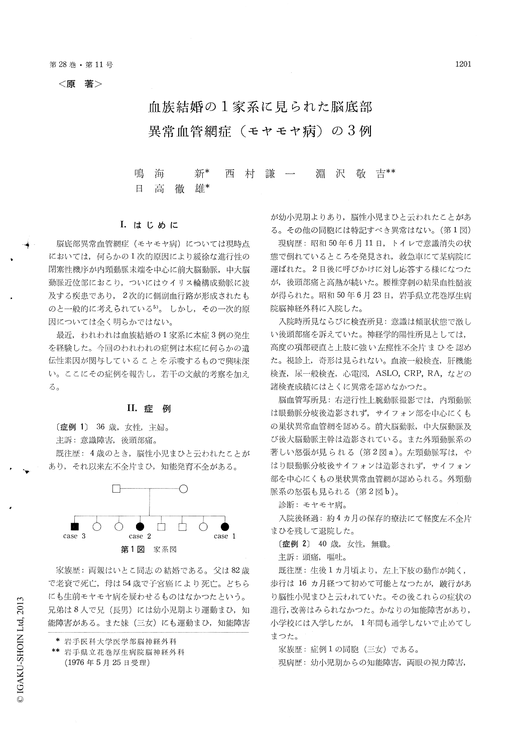
脳底部異常血管網症(モヤモヤ病)については現時点においては,何らかの1次的原因により緩徐な進行性の閉塞性機序が内頸動脈末端を中心に前大脳動脈,中大脳動脈近位部におこり,ついにはウイリス輪構成動脈に波及する疾患であり,2次的に側副血行路が形成されたものと一般的に考えられている5)。しかし,その一次的原因については全く明らかではない。
最近,われわれは血族結婚の1家系に本症3例の発生を経験した。今回のわれわれの症例は本症に何らかの遺伝性素因が関与していることを示唆するもので興味深い。ここにその症例を報告し,若干の文献的考察を加える。
The first case was a 36-year-old woman whose parents were cousins. She was the last of eight siblings. She had suffered from mental retardation and slight motor weakness of the left upper and lower extremities since the age of four. On the 11th of June, 1975, she was found unconscious in the bathroom and admitted to a local hospital. A lumbar puncture was performed there, which re-vealed blood-stained cerebrospinal fluid. She was then transferred to the Hanamaki Kosei Hospital on the 23rd of June. On admission to the hospital, she was still drowsy and showed severe nuchal regidity and left spastic hemiparesis. Cerebral angiograms showed a basal hypervascular area re-sembling a network, and stenosis in both internal carotid arteries at the bifurcation of the ophthalmic artery. Enlarged branches of the external carotid artery were also noted in the angiograms. Under the diagnosis of the Moyamoya disease, she was treated conservatively for 4 months and discharged with remaining mental deterioration and slight left spastic hemiparesis. The second case was her 40-year-old sister. She was the fourth sibling in the family. She also had been suffering from mentalretardation, visual disturbance, and a motor weakness of the left upper and lower extremities since girl-hood. On the 11th of September, 1975, she sudden-ly developed non coma-producing subarachnoid hemorrhage, and was admitted to the Hanamaki Kosei Hospital. The right cerebral angiograms showed a basal vascular network and extremely poor opacification of the main cerebral arteries. Also, enlarged branches of the right external carotid artevy were seen in the angiograms. She was also diagnosed as having the Moyamoya disease and was treated conservatively for one month and discharged. Upon discharge, an examination revealed mental retardation, bilateral myopia, hearing difficulty on the left, and left spastic hemiparesis associated with sensory disturbances to all modalities. The last case was their 60-year old brother. He was the first sibling of the family. He had been suffering from mental retardation, visual disturbance, and a motor weakness of the right upper and lower extremities since boyhood. Recently he began to feel headaches in addition to these neurological symptoms. Left carotid angiograms performed in the Hanamaki Kosei Hospital revealed a basal vascular network, a stenosis of the internal carotid artery at the bifurcation of the ophthalmic artery, and enlarged branches of the external carotid artery. Thus, all three patients showed the typital features of the Moyamoya disease in the cerebral angiograms. The etiology of the Moyamoya disease still remains obscure. These three cases found in an inbred family, however, suggest some genetic factor in the occurrence of the Moyamoya disease.
Copyright © 1976, Igaku-Shoin Ltd. All rights reserved.