Japanese
English
- 有料閲覧
- Abstract 文献概要
- 1ページ目 Look Inside
I.緒言
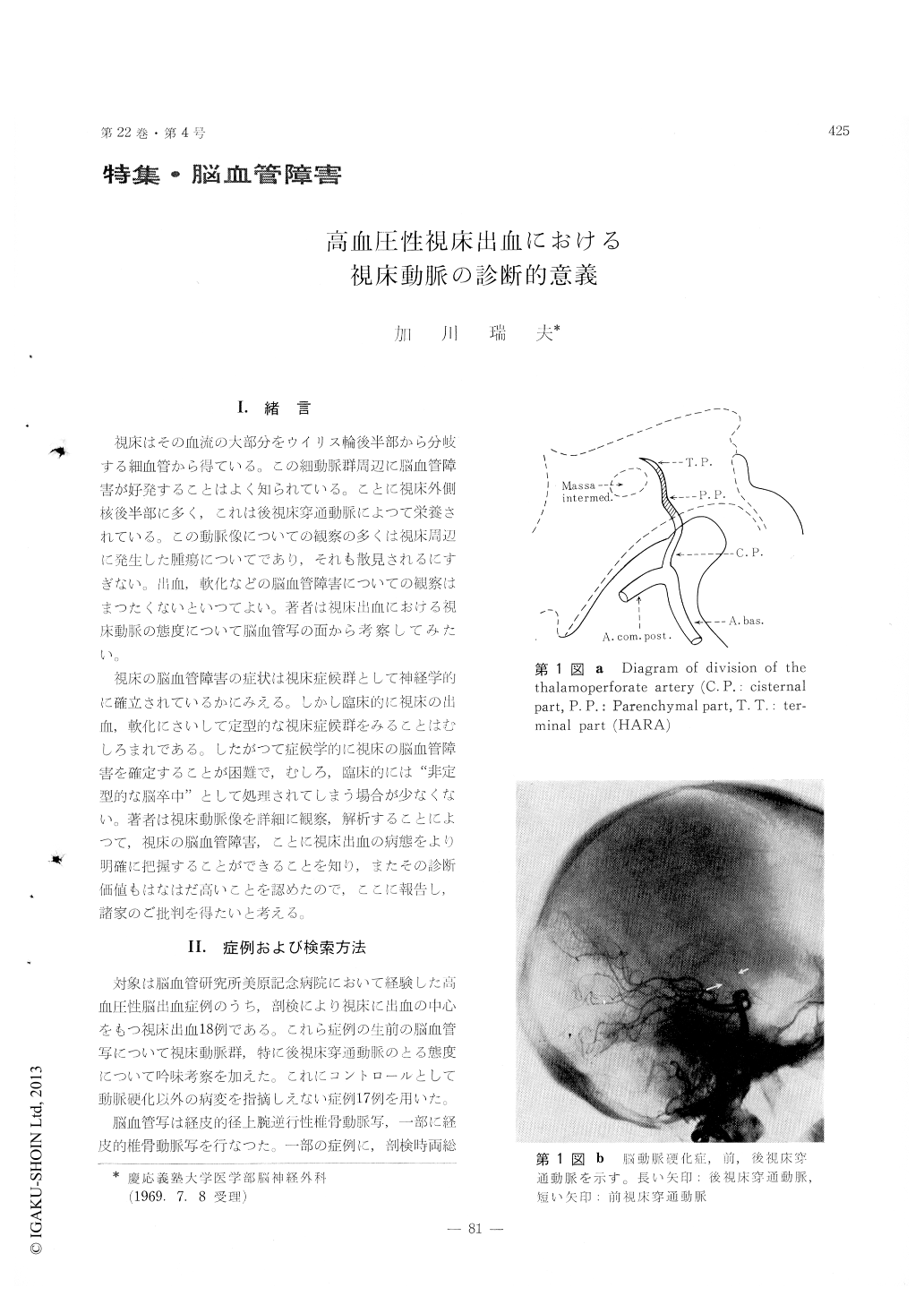
視床はその血流の大部分をウイリス輪後半部から分岐する細血管から得ている。この細動脈群周辺に脳血管障害が好発することはよく知られている。ことに視床外側核後半部に多く,これは後視床穿通動脈によって栄養されている。この動脈像についての観察の多くは視床周辺に発生した腫瘍についてであり,それも散見されるにすぎない。出血,軟化などの脳血管障害についての観察はまつたくないといつてよい。著者は視床出血における視床動脈の態度について脳血管写の面から考察してみたい。
視床の脳血管障害の症状は視床症候群として神経学的に確立されているかにみえる。しかし臨床的に視床の出血,軟化にさいして定型的な視床症候群をみることはむしろまれである。したがって症候学的に視床の脳血管障害を確定することが困難で,むしろ,臨床的には"非定型的な脳卒中"として処理されてしまう場合が少なくない。著者は視床動脈像を詳細に観察,解析することによつて,視床の脳血管障害,ことに視床出血の病態をより明確に把握することができることを知り,またその診断価値もはなはだ高いことを認めたので,ここに報告し,諸家のご批判を得たいと考える。
Cerebrovascular accident of thalamic region occurs mostly due to pathological alteration of tha-lamoperforate arteries.
In this survey, based upon 18 cases with hyper-tensive thalamic hemorrhage, author intended to clarify the angiographic findings of thalamoperforate arteries in hypertensive thalamic hemorrhage, and following conclusions were obtained.
1. Vertebral angiography is one of the most useful diagnostic procedure.
2. The posterior thalamoperforate artery has a great diagnostic value in hypertensive thalamic hemorrhage.
3. The parenchymal part of the posterior thalamo-perforate artery discloses the site and size of he-matoma in the thalamus.

Copyright © 1970, Igaku-Shoin Ltd. All rights reserved.


