Japanese
English
- 有料閲覧
- Abstract 文献概要
- 1ページ目 Look Inside
- 参考文献 Reference
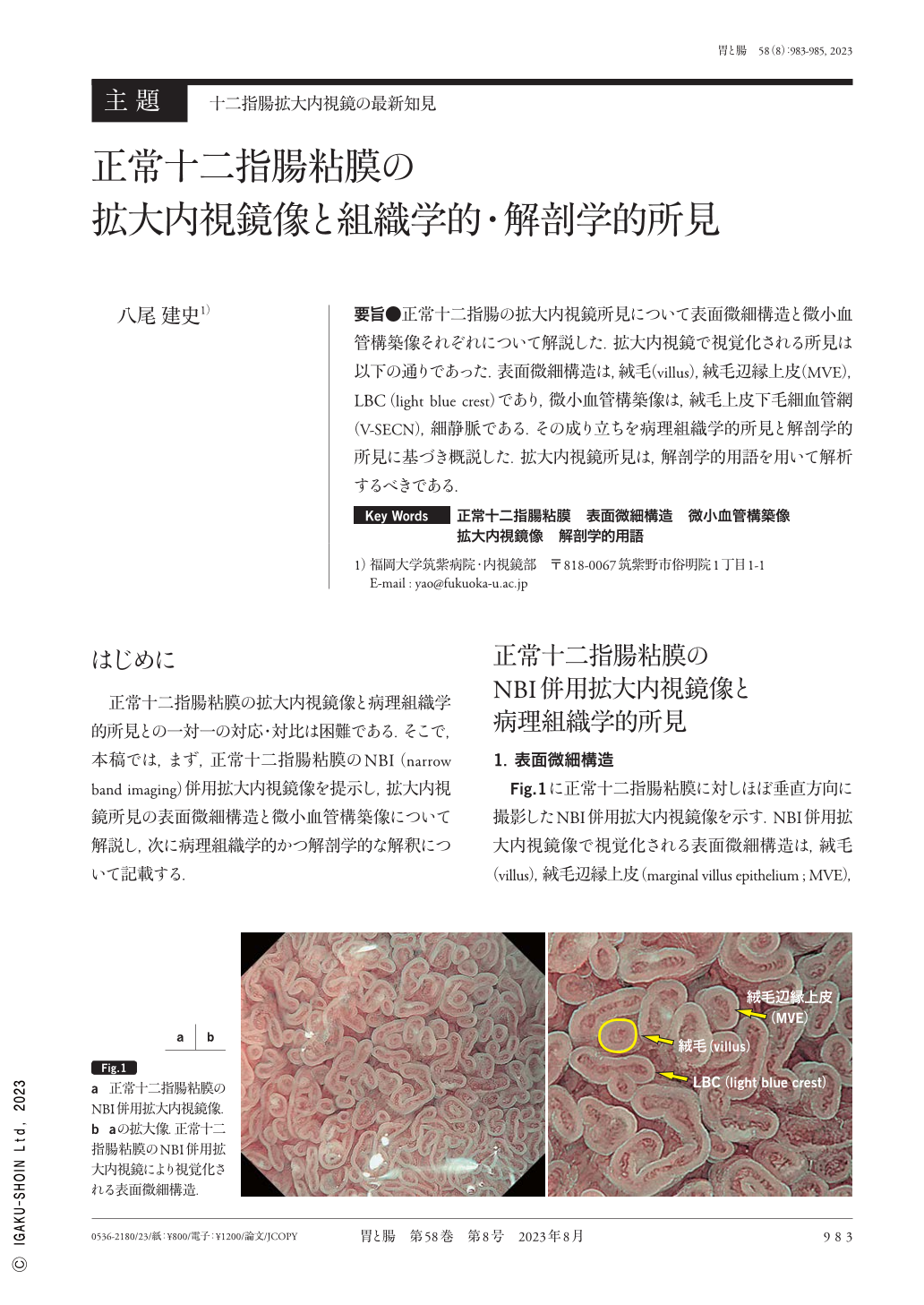
要旨●正常十二指腸の拡大内視鏡所見について表面微細構造と微小血管構築像それぞれについて解説した.拡大内視鏡で視覚化される所見は以下の通りであった.表面微細構造は,絨毛(villus),絨毛辺縁上皮(MVE),LBC(light blue crest)であり,微小血管構築像は,絨毛上皮下毛細血管網(V-SECN),細静脈である.その成り立ちを病理組織学的所見と解剖学的所見に基づき概説した.拡大内視鏡所見は,解剖学的用語を用いて解析するべきである.
Magnified endoscopic findings of normal duodenal mucosa were presented, with a focus on microsurface structure and microvascular architecture. The analysis of microsurface structure revealed the presence of a villus, marginal villus epithelium, and light blue crest. In terms of microvascular architecture, the duodenal mucosa had a villus subepithelial capillary network and venule. The nature of these findings was demonstrated according to the histological and anatomical findings. The magnified endoscopic findings should be described using the aforementioned anatomical terms.

Copyright © 2023, Igaku-Shoin Ltd. All rights reserved.


