Japanese
English
- 有料閲覧
- Abstract 文献概要
- 1ページ目 Look Inside
- 参考文献 Reference
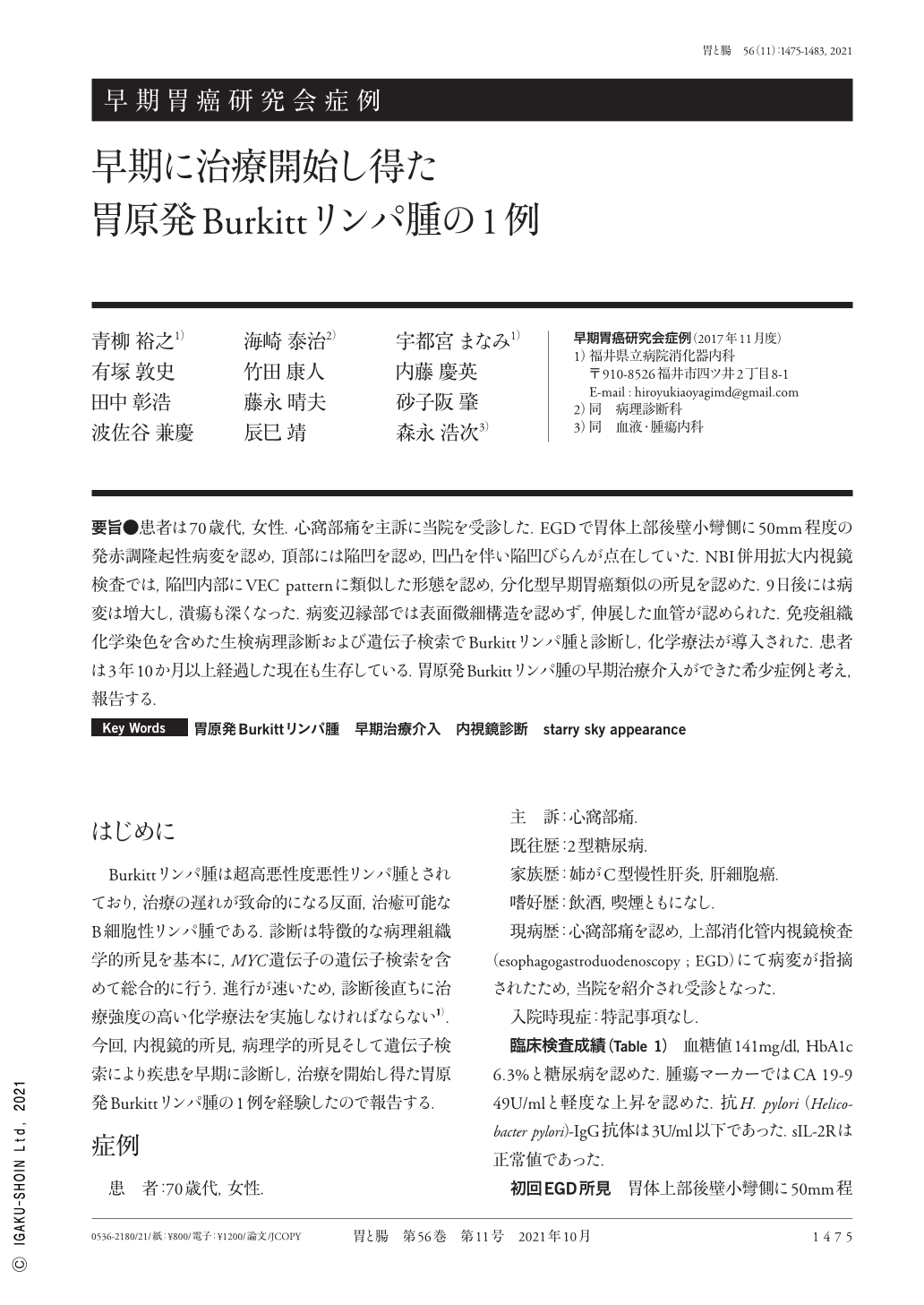
要旨●患者は70歳代,女性.心窩部痛を主訴に当院を受診した.EGDで胃体上部後壁小彎側に50mm程度の発赤調隆起性病変を認め,頂部には陥凹を認め,凹凸を伴い陥凹びらんが点在していた.NBI併用拡大内視鏡検査では,陥凹内部にVEC patternに類似した形態を認め,分化型早期胃癌類似の所見を認めた.9日後には病変は増大し,潰瘍も深くなった.病変辺縁部では表面微細構造を認めず,伸展した血管が認められた.免疫組織化学染色を含めた生検病理診断および遺伝子検索でBurkittリンパ腫と診断し,化学療法が導入された.患者は3年10か月以上経過した現在も生存している.胃原発Burkittリンパ腫の早期治療介入ができた希少症例と考え,報告する.
We report a rare case of BL(Burkitt's lymphoma)of the stomach in a woman in her 70s that was successfully managed by early chemotherapeutic intervention. Esophagogastroduodenoscopy for epigastralgia in our patient revealed a 50mm flat elevated lesion with a shallow depression in the lesser curvature of the upper gastric body under white light imaging. The lesion had a granular surface, a reddish mucosa with erosion, and a shallow ulcer. M-NBI(narrow band imaging with magnification)showed an irregular microsurface and an irregular microvascular pattern with a demarcation line. Importantly, findings in the vessels within epithelial circle were similar to those in the depressed lesion. After 9 days, the lesion increased in size and the ulceration deepened. No surface structure was observed and M-NBI showed the presence of extended blood vessels at the margin of the lesion. Histological analysis of the revealed medium-to-large sized lymphocyte infiltration with “starry sky” macrophages. The lesion was accurately diagnosed as BL based on immunohistochemical analysis and the diagnosis was confirmed by molecular FISH analysis, which detected DNA rearrangements in the MYC gene. The patient was provided intensive multi-agent chemotherapy, including R-hyper CVAD and DA EPOCH-R. Currently, at 3 years and 10 months after chemotherapy initiation, she remains alive and well despite a Dumbbell-like shaped stomach. This case report indicates that gastric BL can be successfully managed if aggressively treated in the early stages.

Copyright © 2021, Igaku-Shoin Ltd. All rights reserved.


