Japanese
English
- 有料閲覧
- Abstract 文献概要
- 1ページ目 Look Inside
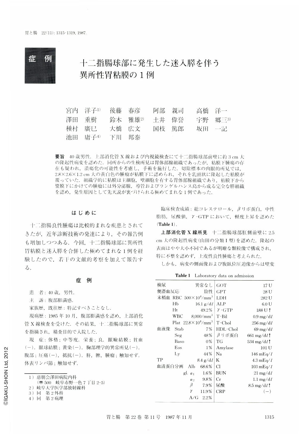
要旨 40歳男性.上部消化管X線および内視鏡検査にて十二指腸球部前壁に約3cm大の隆起性病変を認めた.同所からの生検所見は胃体部腺組織であったが,粘膜下腫瘍の存在も疑われ,潰瘍化の可能性を考慮し,手術を施行した.切除標本の肉眼的所見では,2.8×2.6×1.2cm大の黄白色の腫瘤が粘膜下に認められ,それを乳頭状に隆起した粘膜が覆っていた.組織学的に粘膜は主細胞,壁細胞を有する胃体部腺組織であり,粘膜下から漿膜下にかけての腫瘤には外分泌腺,導管およびランゲルハンス島から成る完全な膵組織を認め,発生原因として先天説が裏づけられる極めてまれな1例であった.
The patient was a 40 year-old man complaining of stomach discomfort for several months. X-ray and endoscopic examinations revealed an elevated lesion, about 3 cm in diameter on the anterior wall of the duodenal bulb. Biopsy specimen showed a fundic gland type gastric mucosa with chief cells and parietal cells. Our clinical diagnosis was that it was a case of benign heterotopic gastric mucosa associated with some submucosal tissue enlargement. Since the patient had gastric distress and the lesion may have producede ulceration, surgical excision was performed. On the surgical specimen, the tumor was 28×26×12 mm in size and covered with papillary swollen mucosa. At the cut section of the specimen, it was found that the tumor was located in the submucosal layer and presented a well demarcated yellowish white mass. Histologically, the papillary swollen mucosa was formed by fundic type gastric mucosa consisting of chief cells and parietal cells. In addition, complete pancreatic tissue consisting of exocrine glands, excretory ducts and islands of Langerhans was disclosed in the layers from submucosa to subserosa. These findings strongly suggest that the etiology of this lesion should be regarded as congenital.

Copyright © 1987, Igaku-Shoin Ltd. All rights reserved.


