Japanese
English
- 有料閲覧
- Abstract 文献概要
- 1ページ目 Look Inside
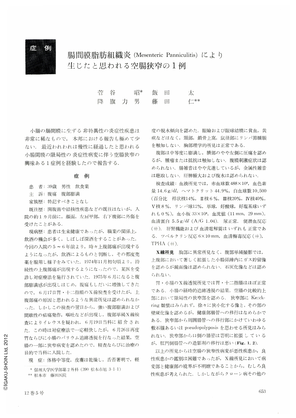
小腸の腸間膜に生ずる非特異性の炎症性疾患は非常に稀なもので,本邦における報告も極めて少ない.最近われわれは慢性に経過したと思われる小腸間膜の限局性の炎症性病変に伴う空腸狭窄の興味ある1症例を経験したので報告する.
A 39-year-old male, who had no tuberculosis or previous operations, had suffered from progressive epigastric pain for about 6 years prior to his admission. He had been conservatively treated by his home doctor since November 1974. As his increasing epigastric pain became accompanied by abdominal distension, he took an upper GI with barium meal on June 17, 1975. Just from the next day he developed severe colicky pain with frequent vomiting and marked abdominal distension. He was immediately referred to our surgical department and treated for a time conservatively. On June 26 the gastrointestinal tract was examined again. It demonstrated a segmental stenotic lesion with gradual tapering in the proximal portion of the jejunum. An absence of Kerckring's folds in the segment and poor transit of the meal to the distal portion of jejunum were shown. On physical examination, his abdomen was moderately distended. There was localized tenderness in the left paraumbilical region but no palpable tumor and muscular rigidity.
Laboratory findings on admission consisted of the following: RBC 4.88 millions, hemoglobin 14.6 gm., hematocrit 44.9%, WBC 10,500, with 88% polymorphonuclears and 12% lymphocytes, blood sedimentation 11 mm, urinalysis w.n.l., fecal occult blood strongly positive, total protein 5.5 gm., with an A/G of 1.04, liver & renal function tests w.n.l., tuberculin test 6×10 mm, serological test for syphilis positive, TPHA (++).
On July 9, segmental resection of the jejunum and jejuno-jejunostomy were performed. Operation findings were: No free fluid and hepatosplenomegaly were encountered. Jejunal stenotic lesion, about 12 cm in length, was located in about one meter distal to the ligament of Treitz. Jejunal mesentery corresponding to the stenotic area showed moderately inflammatory changes with reddish and thickened adipose tissues. Some adjacent organs were firmly adherent to the involved mesentery. The intestinal loop proximal to the stenotic segment was considerably dilated and edematous. All other viscera were grossly normal. In the jejunal mesentery, histologic examination revealed a moderate infiltration of fat by macrophages with occasional apperance of lymphocytes and some degenerative or fibrotic parts. The mucosal surface of the stenotic lesion showed pseudomembranous, hemorrhagic and necrotic change which was limited to the very well-defined area.
The final pathological diagnosis was chronic panniculitis of the mesentery with subsequent pseudomembranous necrotizing enteritis. We concluded that chronic intestinal circulatory disturbance due to the mesenteric panniculitis resulted in necrotizing enteritis with secondary jejunal stenosis.

Copyright © 1977, Igaku-Shoin Ltd. All rights reserved.


