Japanese
English
- 有料閲覧
- Abstract 文献概要
- 1ページ目 Look Inside
Ⅰ.はじめに
胃梅毒は,1834年Andral2)の報告以来かなり多くの報告がなされてきたが,駆梅療法の進歩と共に激減し,最近10年間は,山田10),三方4),R. M. Mitchell5)らの例を散見するのみであった.昨年は,丸山6),春日井3),西浦7),柚木9)らの報告があり,胃疾患診断法の急速な進歩と共に,早期胃癌との鑑別7)9)あるいは早期梅毒に認められる胃粘膜変化6)などの問題を伴って新しく再登場してきた感がある.われわれは,皮膚に梅毒性乾癬を認めた第2期梅毒患者で,胃に多彩な変化を認め,胃癌を疑って胃切除術を施行したが,胃梅毒であった症例を経験したので報告する.
This is a report of gastric syphilis, presumably precocious tertiarioma, found in a man, who was suffering from secondary lues with lichen syphiliticus on his skin and was at the same time found to have very diversified changes in his stomach, later undergoing gastrectomy as gastric cancer was suspected.
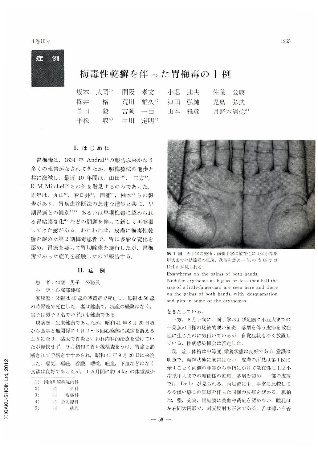
The patient, a 42-year-old man, and dull pain in the epigastrium as his chief complaint. On the palms of his hands and on the soles of his feet were observed here and there desquamation and nodular erythemas as big as or less than half the size of the nail of a little finger. Macroscopically and by histological finding of the biopsied specimen, there changes on the skin were diagnosed as of lichen syphiliticus. Serological reactions for syphilis were: Ogata's test-3 (+), VDRL's slide test-3 (+), flocculation test-3 (+) and quantitative test-320 dls.
At x-ray examination of the stomach, it was found that next to the rigidity and poor distensibility of the gastric walls transversely all the way around extending from the gastric angle to the pyloric antrum, the mucosal folds running from the cardia down to the pylorus was completely obliterated in the pyloric antrum, where irregular-shaped, wide and shallow depressions intermixed with a number of protuberances of various sizes were visualized. Endoscopy revealed irregular, shallow tissue defects and rough unevenness of the mucosa in the pyloric antrum, the whole area of which was tinged with dirty red color. These lesions, of elastic soft consistency, were diagnosed either as of superficial spreading type of gastric cancer of else syphilitic changes.
In the resected specimen, irregular-shaped, wide and shallow tissue defects, sharply demarcated all around against the normal mucosa from the incisura down to the pyloric region, were observed surrounded by intermingling many protrusions of various sizes and by erosions. The lesions as a whole were soft to the touch. Histopathologically it was found the superficial layer had fallen off, and in the submucosal layer was notable cell infiltration in the form of a band (lymphocytes, histiocytes, polymorphic leucocytes, a number of plasma cells and a few Langhans' giant cells). Numerous rejuvenerated blood vessels and mural hypertrophy in some of the blood vessels were also observed. By these findings this case was diagnosed as of gastric syphilis.

Copyright © 1969, Igaku-Shoin Ltd. All rights reserved.


