Japanese
English
- 有料閲覧
- Abstract 文献概要
- 1ページ目 Look Inside
Ⅰ.はじめに
KussmaulがDesormeauxの照明装置に,47cmの長管をつけ,始めて食道内を観察したのは今から丁度100年前である.その後,Killian,Brünings,Jacksonらにより,器械の改良,技術の進歩がなされ,本検査は広く行なわれるようになってきた.
一方,わが国ではKillianに師事された久保教授が,明治40年に始めて食道鏡を使用して摘出した食道異物を報告したが,その後,久保,小野両教授が中心となって,本法の普及に努力されてきた.本学中山教授も,千葉大在任中,側視式,斜視式,逆視式食道鏡を考案,食道噴門部の診断に力をそそいできた.
食道鏡検査は,ごく最近まで耳鼻科領域において,主に治療面を対象として発達してきた.したがって,器械の進歩に優れた点はみられるが,金属の直管を使うという根本原理は変らず,特殊技術として,専ら医師の技術の習熟に負う所大であった.
近年,食道外科の進歩に伴い,一般の内科,外科医の間でも,食道の微細な病変の診断に,食道鏡検査の必要性が高まってきた.それにつれて,挿入の容易な,苦痛の少ない柔軟な食道鏡の出現が強く望まれていた.
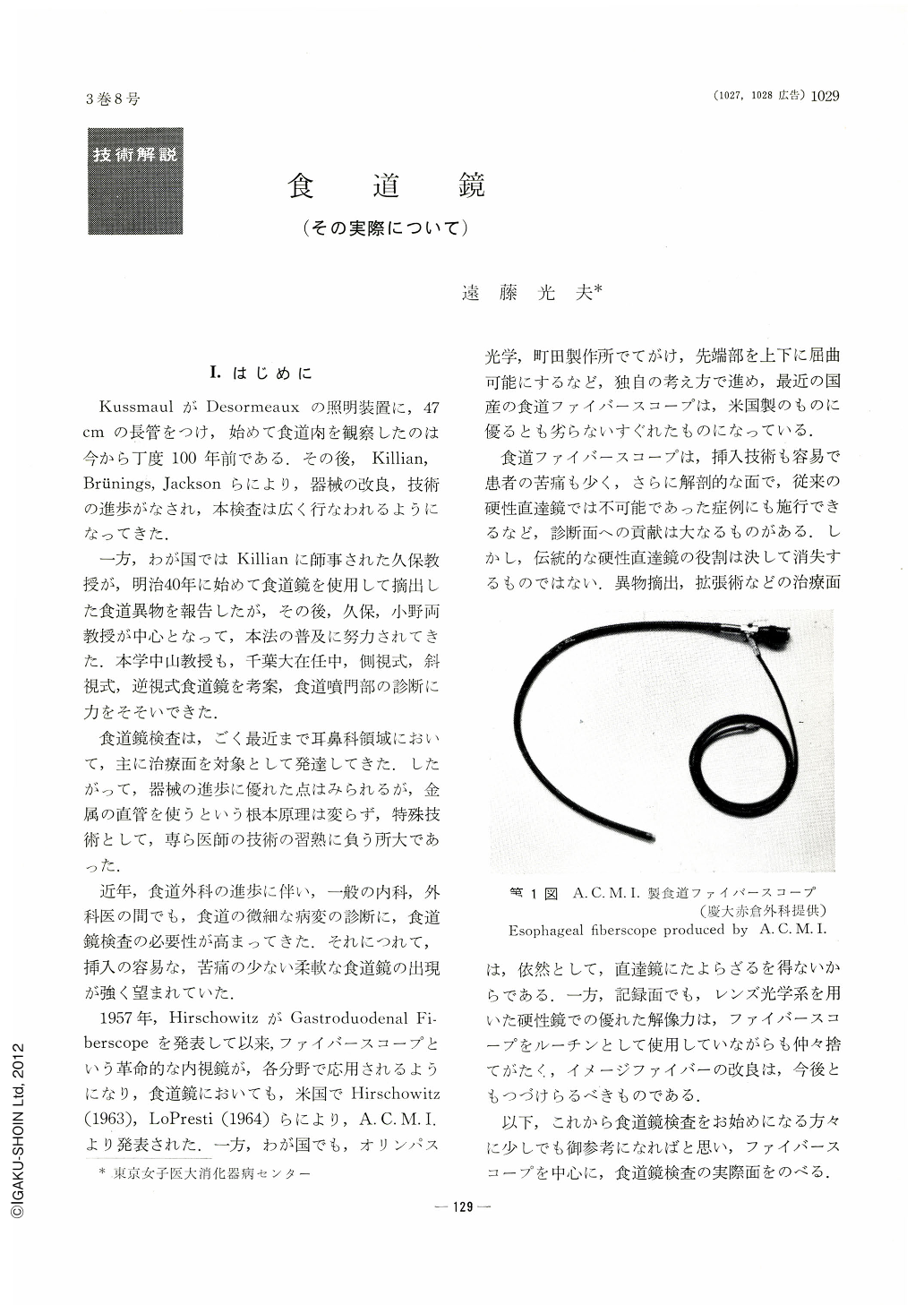
1957年,HirschowitzがGastroduodenal Fiberscopeを発表して以来,ファイバースコープという革命的な内視鏡が,各分野で応用されるようになり,食道鏡においても,米国でHirschowitz(1963),LoPresti(1964)らにより,A. C. M. I.より発表された.一方,わが国でも,オリンパス光学,町田製作所でてがけ,先端部を上下に屈曲可能にするなど,独自の考え方で進め,最近の国産の食道ファイバースコープは,米国製のものに優るとも劣らないすぐれたものになっている.
食道ファイバースコープは,挿入技術も容易で患者の苦痛も少く,さらに解剖的な面で,従来の硬性直達鏡では不可能であった症例にも施行できるなど,診断面への貢献は大なるものがある.しかし,伝統的な硬性直達鏡の役割は決して消失するものではない.異物摘出,拡張術などの治療面は,依然として,直達鏡にたよらざるを得ないからである.一方,記録面でも,レンズ光学系を用いた硬性鏡での優れた解像力は,ファイバースコープをルーチンとして使用していながらも仲々捨てがたく,イメージファイバーの改良は,今後ともつづけらるべきものである.
以下,これから食道鏡検査をお始めになる方々に少しでも御参考になればと思い,ファイバースコープを中心に,食道鏡検査の実際面をのべる.
Esophagoscopy has been developed remarkably since Kussmaul had clone his first inspection of inside of esohagus about 100 years ago. Esophagoscope was made of straight steel tube until recently. It was very dilficult to performe esophagoscopy and this was special technique. Hirschowitz deviced gastoroduodenal fiberscope in 1957, and this new device was applied to esophagoscope, too. In our country, new fiber esophagoscopes were developed by Olympus Optical Company and Machida Company. It is easy to insert new fiberscope and patients' pain is less. By these new fiber scope, the parts of esophagus where hard scope could not see, are able to be seen nowadays. LoPresti's fiber esophagoscope, F. E. S. by Machida Co. and E. F. by Olympus Co. were shown and explanation of the mechanism and how to use were explained. It is necessary to take X-ray photographs of chest and esophagus before the examination. It is also necessary to know whether the patient has heart disease, aneurysm, and anomaly of esophagus. It is the contra indication for hard esophagoscope to do the examination for aneurysm, acute corrosive esophagitis, varix just after the bleeding, tumor of thyroid gland and high grade scoriosis. Even by fiber scope, it is necessary to do the examination with maximum care to these diseases. Five to six hours abstinence from food is necessary before the examination.
Injection for the restriction of excreation and of sedative are administered 30 min. before the examination. Local surface anesthesia is applied to the throat. Usually, examination is performed in the dorsal position but it is possible to do in side or sitting position. The main point and necessary precautions for the insertion were mentioned. Then, how to inspect esophagus was described. Real aspect of the diseases were described. These were esophageal and cardiac cancers, achalasea, varix, anastomosing part between esophagus and stomach or jejunum and the rest stomach after the operation. Method of biopsy was also mentioned. Finally, complications of the examination and its prevention were rles described.

Copyright © 1968, Igaku-Shoin Ltd. All rights reserved.


