Japanese
English
- 有料閲覧
- Abstract 文献概要
- 1ページ目 Look Inside
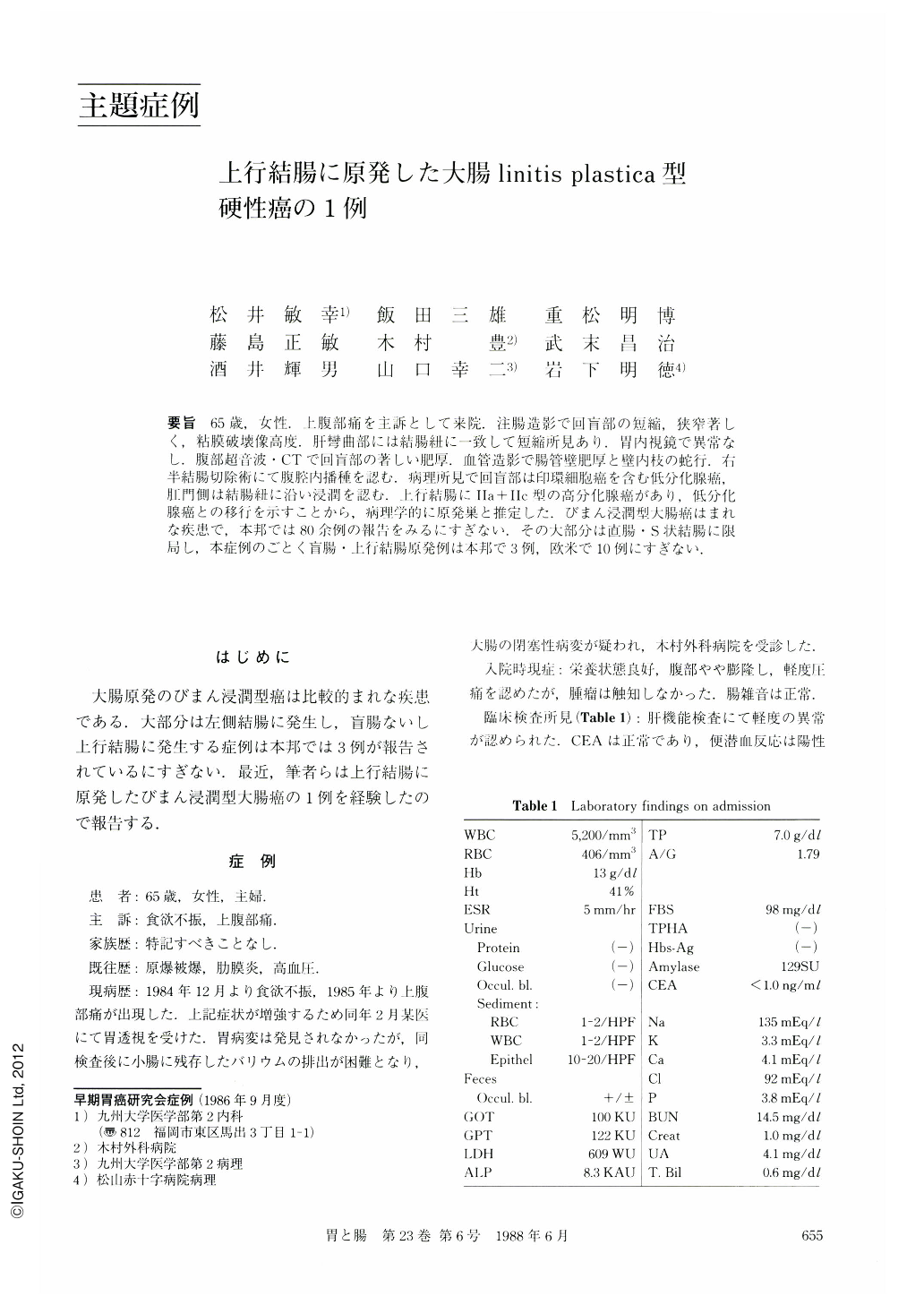
要旨 65歳,女性.上腹部痛を主訴として来院.注腸造影で回盲部の短縮,狭窄著しく,粘膜破壊像高度,肝彎曲部には結腸紐に一致して短縮所見あり.胃内視鏡で異常なし.腹部超音波・CTで回盲部の著しい肥厚.血管造影で腸管壁肥厚と壁内枝の蛇行.右半結腸切除術にて腹腔内播種を認む.病理所見で回盲部は印環細胞癌を含む低分化腺癌,肛門側は結腸紐に沿い浸潤を認む.上行結腸にⅡa+Ⅱc型の高分化腺癌があり,低分化腺癌との移行を示すことから,病理学的に原発巣と推定した.びまん浸潤型大腸癌はまれな疾患で,本邦では80余例の報告をみるにすぎない.その大部分は直腸・S状結腸に限局し,本症例のごとく盲腸・上行結腸原発例は本邦で3例,欧米で10例にすぎない.
We present a case of diffusely infiltrating type of colonic carcinoma. The patient was a 65-year-old woman complaining of upper abdominal pain. Barium enema examination demonstrated an irregularly demarcated narrowing and shortening of the cecum and the ascending colon (Figs. 1 and 2). Endoscopically, mucosal fold convergencies were observed in the transverse colon (Figs. 4 and 5). At operation, peritoneal dissemination was found but a right hemicolectomy was performed. Macroscopically, the resected specimen contained an annular narrowing with prominent thickening of the wall In the ascending colon a Ⅱa + Ⅱc-like lesion measuring 1.2×1.2 cm was observed (Fig. 6). Histologically, this polypoid lesion was well differentiated adenocarcinoma with gradual transition to poorly differentiated adenocarcinoma in the surrounding area (Fig. 7). Thus, it was strongly suggested that this polypoid lesion was the primary lesion. In other parts, there was a massive and scirrhous infiltration of poorly differentiated adenocarcinoma with marked lymphatic permeation (Fig. 7).
Diffusely infiltrative carcinoma of the colon is a rare disorder, especially in the right side of the colon.
Radiographically, this must be differentiated from ischemic colitis or Crohn's disease.

Copyright © 1988, Igaku-Shoin Ltd. All rights reserved.


