Japanese
English
- 有料閲覧
- Abstract 文献概要
- 1ページ目 Look Inside
骨髄以外に発生する形質細胞腫は,髄外性形質細胞腫(extramedullary plasmacytoma)と呼ばれ,その好発部位は上気道,口腔および眼瞼結膜であり,全体の80%を占める.しかし,胃に発生する形質細胞腫は極めて少なく,現在まで欧米で60例余,本邦では11例にすぎない.筆者らは最近monoclonal gammopathyを伴った胃原発性の形質細胞腫を経験したので,免疫学的な検索を加えて報告する.
Extramedullary plasmacytoma occurring in the stomach is exceedingly rare and its incidence estimates about 5% out of those of extramedullary plasmacytoma. Although more than 60 cases of gastric plasmacytomas were reported in the literature, few detailed immunological studies have been carried out In this paper we report a case of gastric plasmacytoma which was extensively examined by morphological and immunological studies.
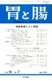
A49-year-old man was first seen because of occational dizziness, loss of appetite and melena of two months' duration. The diagnosis of advanced gastric carcinoma was made by workup of radiologic and endoscopic examinations and he was referred to the Cancer Research Institute Hospital of Kanazawa University for surgery. On admission, there were no particular abnormalities except slight anemia on physical examination. Radiologic and endoscopic examinations revealed generalized giant folds from upper part of fornix downward to mid-body, associated with multiple ulcerations along the running of irregularly enlarged mucosal folds. Histologic examination of biopsies disclosed sheets of malignant cells similar to atypical plasma cells. On February 27, 1979 a total gastrectomy with splenectomy and distal pancreatectomy was performed. Gross appearance of resected stomach showed irregularly enlarged and tortuous folds on the anterior of body measuring 11.5×16.5cm. Histologically the tumor was composed of atypical plasma cells characterized by eccentric nuclei and pyroniophilic cytoplasmas, extending into the entire thickness of the wall. Surgical margins appeared clear. No lymph node involvement was found. As an incidental finding, there was a separate small focus of superficial gastric adenocarcinoma (Ⅱa+Ⅱc type) infiltrating the submucosa in the prepyloric region. Although lymphatic and vascular invasions of carcinoma were slightly present, there was no metastases to lymph nodes or to the liver.
Because the diagnosis of gastric plasmacytoma was strongly suggested by histological examination, the patient's serum was analyzed by means of immunoelectrophoresis. Agar immunoelectrophoresis was performed in the gamma zone M protein which reacted specifically with anti-IgG and anti-lambda antisera. Hewever, agar electrophoresis and immunofixation electrophoresis with anti-lgG and with lambda antisera revealed unusual subbands of M component. lmmunohistochemical studies with peroxidase anti-peroxidase complex (PAP) demonstrated that the tumor cells contained IgG (λ) immunoglobulin. Postoperative course was uneventful and he remained asymptomatic for eight months after gastrectomy when recurrent tumor in the left abdomen and a metastatic lesion in the right orbit were found. At the same time left pleural effusion and mediatinal widening were noted. Cytology examination of the fluid showed atypical plasma cells which were positive for FITCconjugated anti-IgG and anti-lambda. It was noteworthy that the monoclonal immunoglobulin increased in close correlation with recurrent tumor growth. Moreover, lambda type Bence Jones which was previously not seen until August of 1979 was detected with recurrent tumor. The bone survey and bone marrow puncture were unremarkable. The patient died of recurrent tumor and bronchopneumonia 13 months after surgery.
Copyright © 1981, Igaku-Shoin Ltd. All rights reserved.