Japanese
English
- 有料閲覧
- Abstract 文献概要
- 1ページ目 Look Inside
- サイト内被引用 Cited by
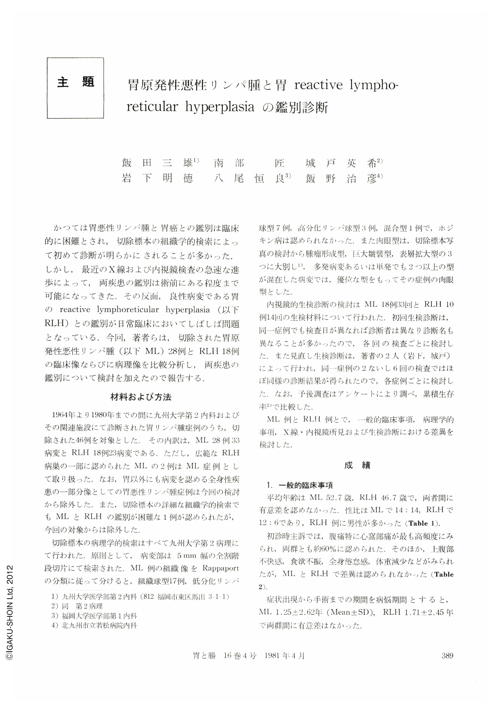
かつては胃悪性リンパ腫と胃癌との鑑別は臨床的に困難とされ,切除標本の組織学的検索によって初めて診断が明らかにされることが多かった.しかし,最近のX線および内視鏡検査の急速な進歩によって,両疾患の鑑別は術前にある程度まで可能になってきた.その反面,良性病変である胃のreactive lymphoreticular hyperplasia(以下RLH)との鑑別が日常臨床においてしばしば問題となっている.今回,著者らは,切除された胃原発性悪性リンパ腫(以下ML)28例とRLH18例の臨床像ならびに病理像を比較分析し,両疾患の鑑別について検討を加えたので報告する.
Analyzing resected 28 cases of primary malignant lymphoma of the stomach (ML) and 18 cases of reactive lymphoreticular hyperplasia of the stomach (RLH), we compared and discussed their differences in clinical feature, pathological findings, radiographic and endoscopic findings as well as biopsy diagnosis between the two diseases.
The results were as follows:
1) Regarding age, sex, chief complaints and illperiod, there were no difference between them. The cumulative survival rate during post-operative five years was 65% in ML and 93% in RLH, but no significant difference was noted.
2) Concerning a location of their lesions, a circumferential lesion involving greater and lesser curvature as well as anterior and posterior wall of the stomach was seen in 3% of ML but in 26% of RLH which was significantly high.
3) By analyzing photographic feature of the resected specimens, we classified them into three macroscopic types―namely mass forming type, giant rugal type and superficial spreading type. The mass forming type was seen in 19 cases (68%) of ML and only one case (6%) of RLH, and it was significantly high in ML.
4) Two cases of the giant rugal type were found in both ML and RLH, however by measuring resected specimen and radiographic picture, it was found that the rugae were bigger in ML and their maximal width was more than 10 mm.
5) Superficial spreading type was seen in seven cases (30%) of ML and 15 cases (83%) of RLH and this type was most difficult to be differentiated. It was also noted that Ⅱa-like lesion or slightly elevated lesion was more frequently seen in ML. Furthermore, the elevation was more prominent at the border rather than its center.
6) Diagnosis of ML was definitely made only in 9% and suspected in 21% by initial biopsy evaluation but after re-evaluating the biopsy specimen, the diagnosis was definitely made in 50% and suspected in 33%.
7) Considering the above results, we want to emphasize that even if the biopsy was initially interpreted as benign, a diagnosis of ML should be suspected in tumor forming lesion, giant rugae with their width greater than 10 mm and superficial spreading lesion with marginal or inner elevation, and further biopsies and re-evalution by well trained pathologist should be carried out for such a lesion.

Copyright © 1981, Igaku-Shoin Ltd. All rights reserved.


