Japanese
English
- 有料閲覧
- Abstract 文献概要
- 1ページ目 Look Inside
- サイト内被引用 Cited by
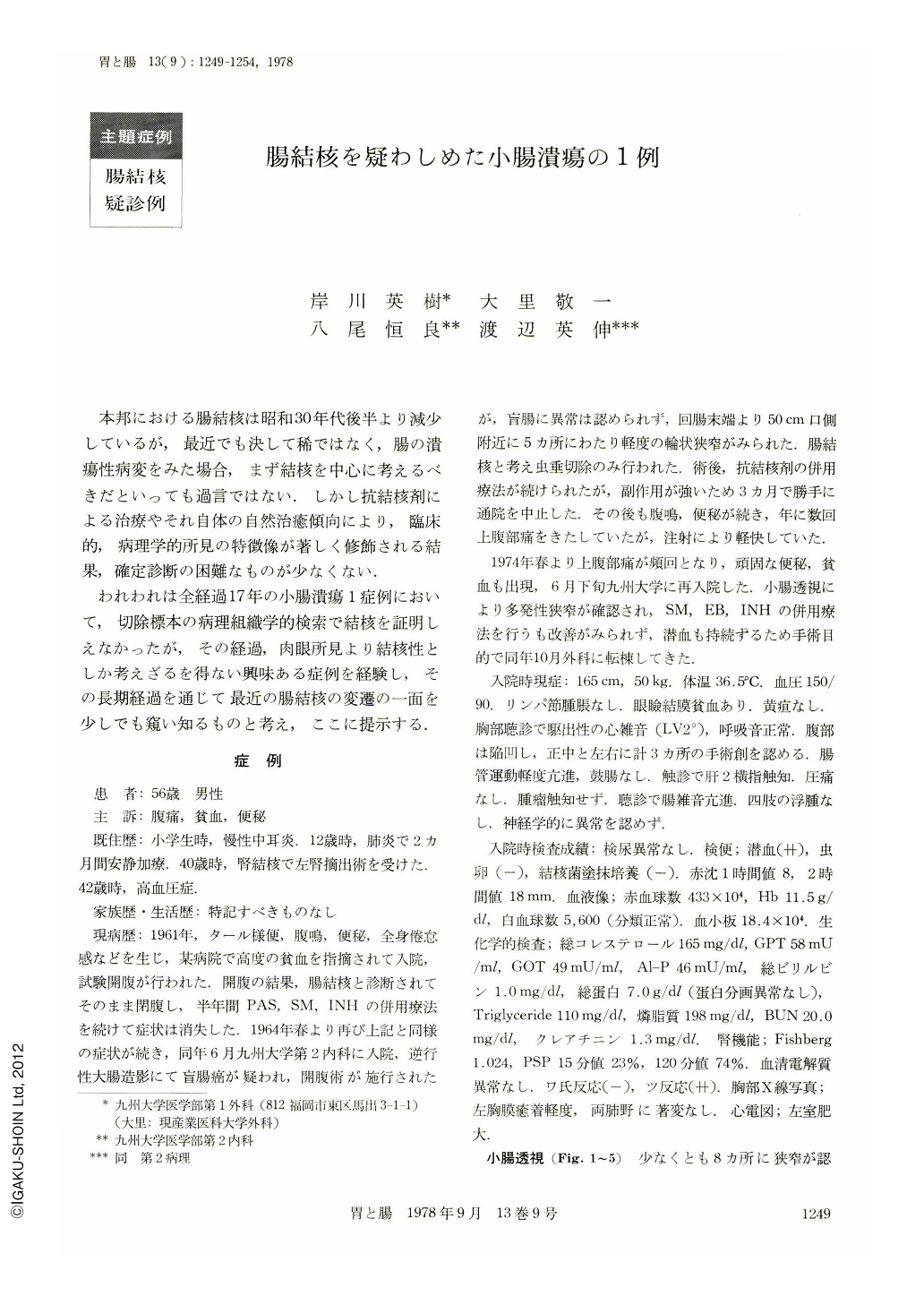
本邦における腸結核は昭和30年代後半より減少しているが,最近でも決して稀ではなく,腸の潰瘍性病変をみた場合,まず結核を中心に考えるべきだといっても過言ではない.しかし抗結核剤による治療やそれ自体の自然治癒傾向により,臨床的,病理学的所見の特徴像が著しく修飾される結果,確定診断の困難なものが少なくない.
われわれは全経過17年の小腸潰瘍1症例において,切除標本の病理組織学的検索で結核を証明しえなかったが,その経過,肉眼所見より結核性としか考えざるを得ない興味ある症例を経験し,その長期経過を通じて最近の腸結核の変遷の一面を少しでも窺い知るものと考え,ここに提示する.
症 例
患 者:56歳 男性
主 訴:腹痛,貧血,便秘
既住歴:小学生時,慢性中耳炎.12歳時,肺炎で2カ月間安静加療,40歳時,腎結核で左腎摘出術を受けた.42歳時,高血圧症.
家族歴・生活歴:特記すべきものなし
現病歴:1961年,タール様便,腹鳴,便秘,全身倦怠感などを生じ,某病院で高度の貧血を指摘されて入院,試験開腹が行われた.開腹の結果,腸結核と診断されてそのまま閉腹し,半年間PAS,SM,INHの併用療法を続けて症状は消失した.1964年春より再び上記と同様の症状が続き,同年6月九州大学第2内科に入院,逆行性大腸造影にて盲腸癌が疑われ,開腹術が施行されたが,盲腸に異常は認められず,回腸末端より50cm口側附近に5ヵ所にわたり軽度の輪状狭窄がみられた.腸結核と考え虫垂切除のみ行われた.術後,抗結核剤の併用療法が続けられたが,副作用が強いため3カ月で勝手に通院を中止した.その後も腹鳴,便秘が続き,年に数回上腹部痛をきたしていたが,注射により軽快していた.
1974年春より上腹部痛が頻回となり,頑固な便秘,貧血も出現,6月下旬九州大学に再入院した.小腸透視により多発性狭窄が確認され,SM,EB,INHの併用療法を行うも改善がみられず,潜血も持続するため手術目的で同年10月外科に転棟してきた.
A 56-year-old man was admitted to our clinic with a long history of recurrent abdominal pain, anemia, gurgling sounds and constipation.
He had a history of left nephrectomy for renal tuberculosis in 1960.
His first abdominal symptoms occurred in 1961, when he was admitted to a local hospital due to tarry stool, gurgling sounds, constipation and general fatigue. On explorative laparotomy he was diagnosed as having intestinal tuberculosis and was on antituberculous treatment for six months with good result. Since then he was free from symtoms until 1964, when he was admitted to our clinic due to the same complaints. On exploration there were five encircling strictures at the distal ileum 50 cm away from the ileum end. He was treated again by the antituberculosis drugs for three months. After that time he improved in health except occasional slight abdominal pain. In 1974 he was readmitted to our clinic due to frequent abdominal pain and general fatigue. On admission he had anemia and hypoproteinemia. Roentgenographic examination showed multiple strictures of the small intestine. His Mantoux reaction was positive but continuous fecal culture for tubercle bacillus was always negative. Occult blood of stool was always positive. Chest X-ray films revealed no abnormal shadow. As his symptoms were not improved by antituberculous treatment, operation (multiple resections with end-to-end anastomoses) was carried out. The resected specimens showed multiple linear ulcers from the distal jejunum to the proximal ileum, and two pseudoenteloliths coexisted with these ulcers.
The histological specimens showed chronic non-specific ulcers (Ul-Ⅱ~Ⅳ), two ulcers being open, and two atrophic non-caseating granulomas in the submucosa of one of 71 sections. Mesenteric lymph nodes revealed only chronic non-specific lymph-adenitis.
Postoperative course was uneventful and he has been doing well since discharge.
In this case tubercle bacilli or microscopic tuberculous lesions were not recognized but clinical course and macroscopic appearance strongly suggest tuberculosis of the small intestine.

Copyright © 1978, Igaku-Shoin Ltd. All rights reserved.


