Japanese
English
- 有料閲覧
- Abstract 文献概要
- 1ページ目 Look Inside
- サイト内被引用 Cited by
linitis plastica型癌は種々の肉眼型の胃癌のうちでもっとも予後の悪い肉眼型とされているが,その概念や定義については諸家の間でいささか見解の相違がある.組織学的に見られる著明な線維形成をEppingerらは浮腫部に見られる漏出血漿成分による漿液性炎症の結果とし,佐野らはその過程を水溶性コラーゲンが不溶性コラーゲンに変化するためとしている.一方,乳腺の硬性癌では癌細胞が結合織線維を産生するという報告がある1).
linitis plastica型胃癌は広範に浸潤しているためにその原発巣の検索は従来十分になされていなかった.Saphir & Parker(1943)は幽門前庭部に原発する癌が胃体部へびまん性浸潤をしてlinitis plastica型癌が形成されることが多いと述べている.近年に至り経過観察例が報告され,原発巣の部位や形態が解明されつつある10).佐野らはⅡc型癌の一部に,また中村らは胃底腺粘膜に発生した癌に原発を求めている.
We made a comparative study on 42 cases of linitis plastica carcinoma and 23 cases of localized diffuse carcinoma.
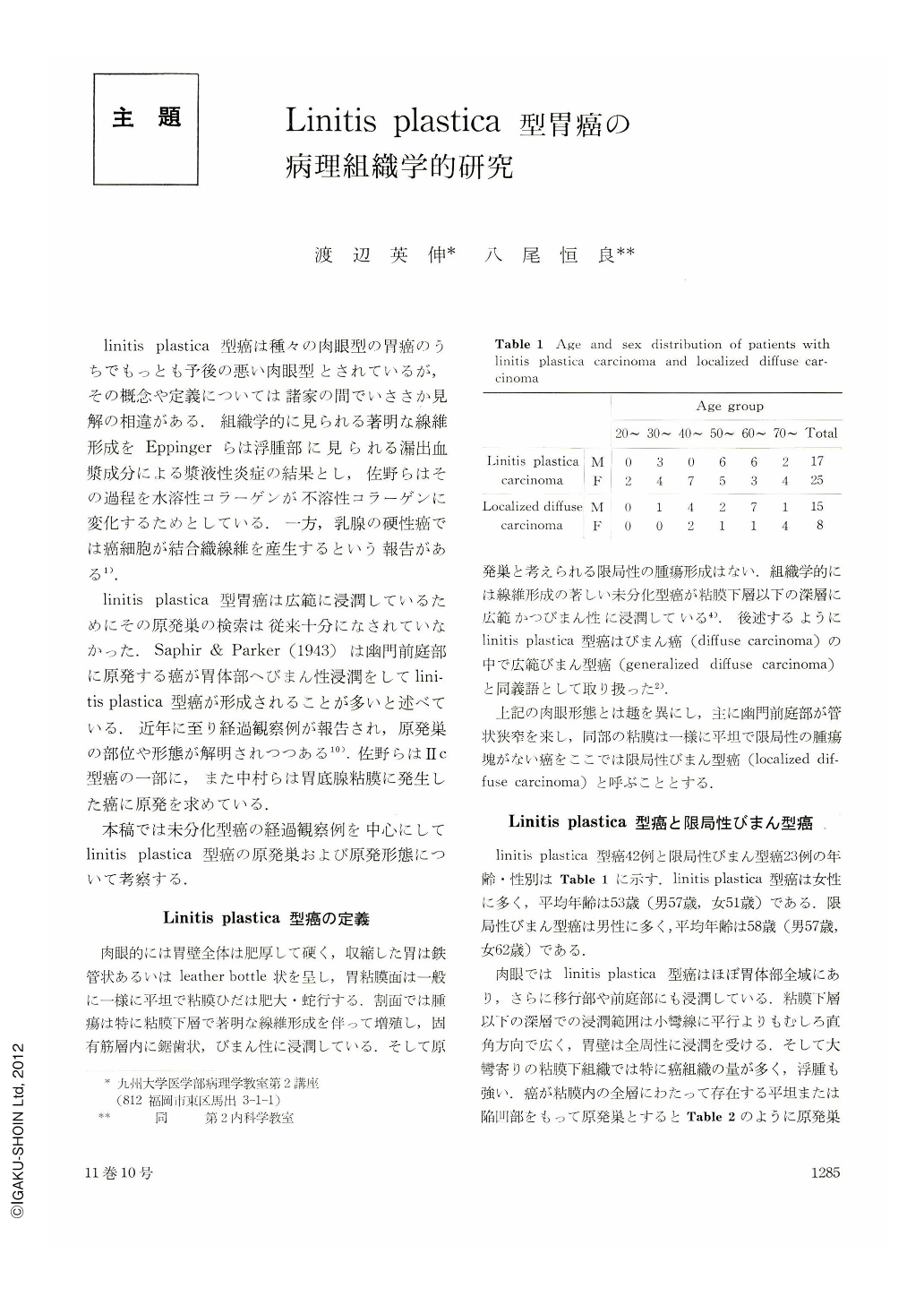
There are some differences between them: the site of a primary focus; ratio of the size of the largest dimensions between the mucosal spreading of a primary focus and its speading in the submucosa and further deep layers, etc., but no significant differences were seen in histologic type of carcinoma and prognosis (Tables 1, 2 & 3).
The macroscopic difference between them seems only to be due to difference in the primary site of the carcinoma: linitis plastica carcinoma arising in fundic glandular area and junctional zone against localized diffuse carcinoma arising in the pyloric glandular area and junctional zone.
Therefore they should be classified into one type of gastric carcinoma, i. e., diffuse carcinoma, and it sholud be subdivided into linitis plastica carcinoma (generalized diffuse carcinoma) and localized diffuse carcinoma.
We searched for the macroscopic type of a primary focus in linitis plastica carcinoma, using 5 undifferentiated carcinomas developed into linitis plastica type and 29 undifferentiated carcinoma developed into other types during observation.
Out of the former five cases, four were estimated on the first X-ray examination to have been a IIc-type (or like advances) carcinoma and one a small advanced scirrhous carcinoma with a relatively small ulcer as primary macroscopic pattern.
Eighteen cases followed a course of a malignant cycle and did not disclose any visible change in the tumor size. These cases were associated with a dense fibrosis surrounding the advancing margin of the tumor, which was never seen in linitis plastica carcinoma.
In a group showing an increase in tumor size during observation, a localized mass was seen in five cases, of which three were an ulceration infiltrative carcinoma and two a carcinoma with lymphoid stroma.
Three cases of the remainder six cases without peptic ulcer manifested markedly increased rigidity of the gastric wall on the X-ray examination done about one to four years later. This finding was quite similar to that of the first X-ray in cases developed into linitis plastica carcinoma. The resected stomach showed a IIc-like advanced carcinoma, histologically reminding one of a small diffuse carcinoma.
As results, we can conclude that linitis plastica carcinoma has a primary focus in the fundic glandular mucosa or junctional mucosa and that a primary focus is a IIc-type undifferentiated carcinoma without peptic ulcer; even if the ulcer is present, it is relatively small as compared with the tumor tissue existing in the submucosa and further deep layers.
Copyright © 1976, Igaku-Shoin Ltd. All rights reserved.