Japanese
English
- 有料閲覧
- Abstract 文献概要
- 1ページ目 Look Inside
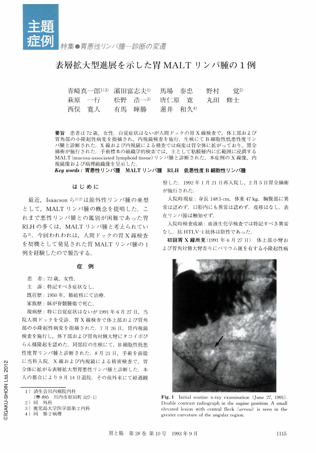
要旨 患者は72歳,女性.自覚症状はないが人間ドックの胃X線検査で,体上部および胃角部の小隆起性病変を指摘され,内視鏡検査を施行.生検にてB細胞性低悪性度リンパ腫と診断された.X線および内視鏡による精査では病変は胃全体に拡がっており,胃全摘術が施行された.手術標本の組織学的検索では,主として粘膜層内に広範囲に浸潤するMALT(mucosa-assoclated lymphoid tissue)リンパ腫と診断された.本症例のX線像,内視鏡像および病理組織像を呈示した.
Endoscopic examination was carried out in a 72-year-old female for small elevated lesions in the upper body and angular region of her stomach which were indicated by medical checkup-gastric roentgenography. She had no subjective symptom. Low-grade malignant B-cell lymphoma of the stomach was diagnosed from the histology of the biopsied specimens.
The detailed x-ray examination showed abnormal areae gastricae extending to the whole stomach. The abnormality of areae gastricae contained irregularity in size and form, and disorders of direction and arrangement. Sulcus-like erosions surrounding the individual areae gastricae were sporadically observed. Endoscopic examination showed unevenness of the mucosa distributing on the whole stomach. The mucosa was composed of variously sized and smooth-surfaced granules with sporadically distributed sulcus-like erosions which was revealed by dye spraying method.
We then diagnosed as gastric malignant lymphoma of superficial spreading type. Total gastrectomy was subsequently performed for the patient.
Examination of the whole stomach resected in the serial sections showed that malignant lymphoma (ML) cells revealed infiltrative proliferation with sporadic or diffuse growth pattern in the mucosa of the whole stomach and involved focally the submucosal layer. No metastasis of the ML was seen in the regional lymph nodes examined. The ML cells were small lymphocytic, comprising cytologically so-called centrocytoid cells, monocytoid cells and plasmacytoid cells. These ML cells formed lymphoepithelial lesions in the mucosa. Paraffin-immunohistochemically the ML cells were B cells and revealed differentiation to plasma cells having monoclonal cytoplasmic immunoglobulin. Thus, this case was diagnosed as mucosa-associated lymphoid tissue (MALT)-type malignant lymphoma involving almost whole mucosa of the stomach.
Copyright © 1993, Igaku-Shoin Ltd. All rights reserved.