Japanese
English
- 有料閲覧
- Abstract 文献概要
- 1ページ目 Look Inside
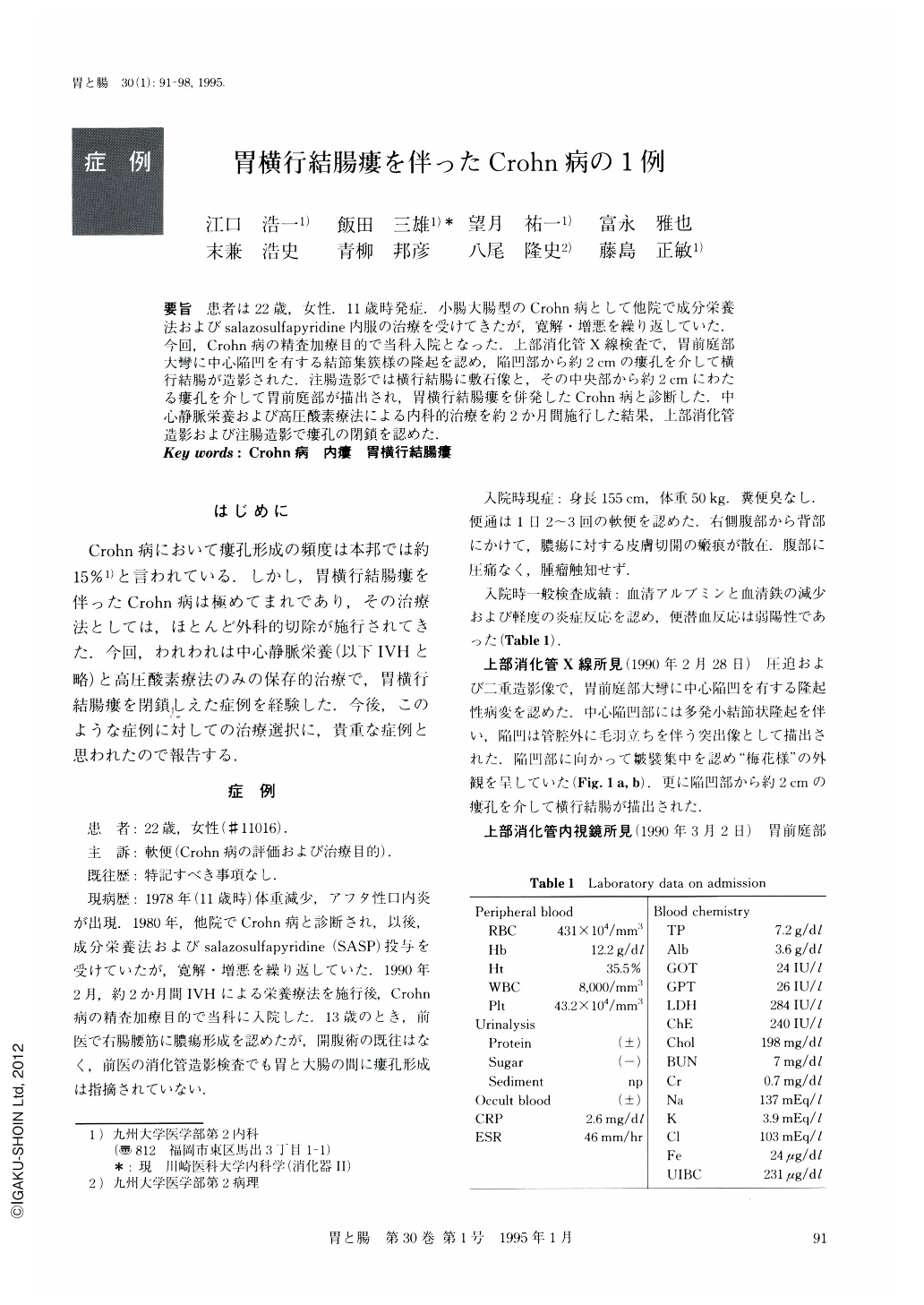
要旨 患者は22歳,女性.11歳時発症.小腸大腸型のCrohn病として他院で成分栄養法およびsalazosulfapyridine内服の治療を受けてきたが,寛解・増悪を繰り返していた.今回,Crohn病の精査加療目的で当科入院となった.上部消化管X線検査で,胃前庭部大彎に中心陥凹を有する結節集簇様の隆起を認め,陥凹部から約2cmの瘻孔を介して横行結腸が造影された.注腸造影では横行結腸に敷石像と,その中央部から約2cmにわたる瘻孔を介して胃前庭部が描出され,胃横行結腸痩を併発したCrohn病と診断した.中心静脈栄養および高圧酸素療法による内科的治療を約2か月間施行した結果,上部消化管造影および注腸造影で瘻孔の閉鎖を認めた.
A 22-year-old woman was admitted to our hospital for further examination of Crohn's disease. She had been treated with elemental diet and salazosulfapyridine after diagnosis of Crohn's disease of the ileocolitis type. An upper gastrointestinal series of investigations demonstrated an elevated lesion with a central depression on the greater curvature of the antrum, and revealed a fistulous tract (about 1.5 cm in length), communicating with the midtransverse colon. On barium enema radiography, cobblestone appearance, eccentric narrowing, and a fistula from the transverse colon to the stomach were also seen. Based on these findings, this case was considered to be Crohn's disease complicating gastrocolic fistula. She was successfully treated with intravenous hyperalimentation and hyperbaric oxygen for about two months.

Copyright © 1995, Igaku-Shoin Ltd. All rights reserved.


