Japanese
English
- 有料閲覧
- Abstract 文献概要
- 1ページ目 Look Inside
- サイト内被引用 Cited by
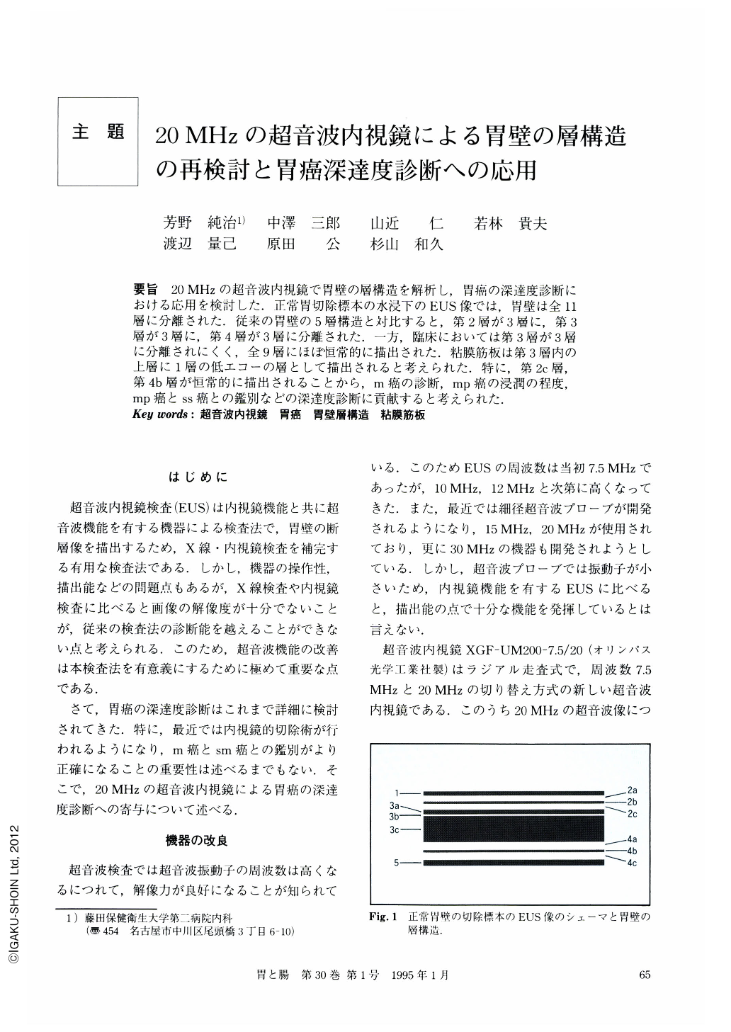
要旨 20MHzの超音波内視鏡で胃壁の層構造を解析し,胃癌の深達度診断における応用を検討した.正常胃切除標本の水浸下のEUS像では,胃壁は全11層に分離された.従来の胃壁の5層構造と対比すると,第2層が3層に,第3層が3層に,第4層が3層に分離された.一方,臨床においては第3層が3層に分離されにくく,全9層にほぼ恒常的に描出された.粘膜筋板は第3層内の上層に1層の低エコーの層として描出されると考えられた.特に,第2c層,第4b層が恒常的に描出されることから,m癌の診断,mp癌の浸潤の程度,mp癌とss癌との鑑別などの深達度診断に貢献すると考えられた.
We studied the mural structure of the stomach by 20MHz-ultrasonographic endoscope and its application for the diagnosis of gastric cancerous invasion. The walls of resected stomachs were observed as 11 layers in a water tank by the new apparatus. The second layer, the third layer, and the fourth layer in the conventional five-layered structure were divided into three layers respectively. On the other hand, clinically, the walls of stomachs revealed nine layers, because the third layer was not divided into three layers in most cases. It was speculated that the muscularis mucosae corresponds to the 3b layer which was observed as a hypoechoic layer in the upper part of the conventional third layer. Because the 2c layer (the lower layer in the conventional second layer) and the 4b layer (the middle layer in the conventional fourth layer) were able to be observed clearly, diagnosis of the depth of gastric cancerous invasion is expected to be improved through the use of such. apparatuses as the 20MHz-ultrasonographic endoscope.

Copyright © 1995, Igaku-Shoin Ltd. All rights reserved.


