Japanese
English
- 有料閲覧
- Abstract 文献概要
- 1ページ目 Look Inside
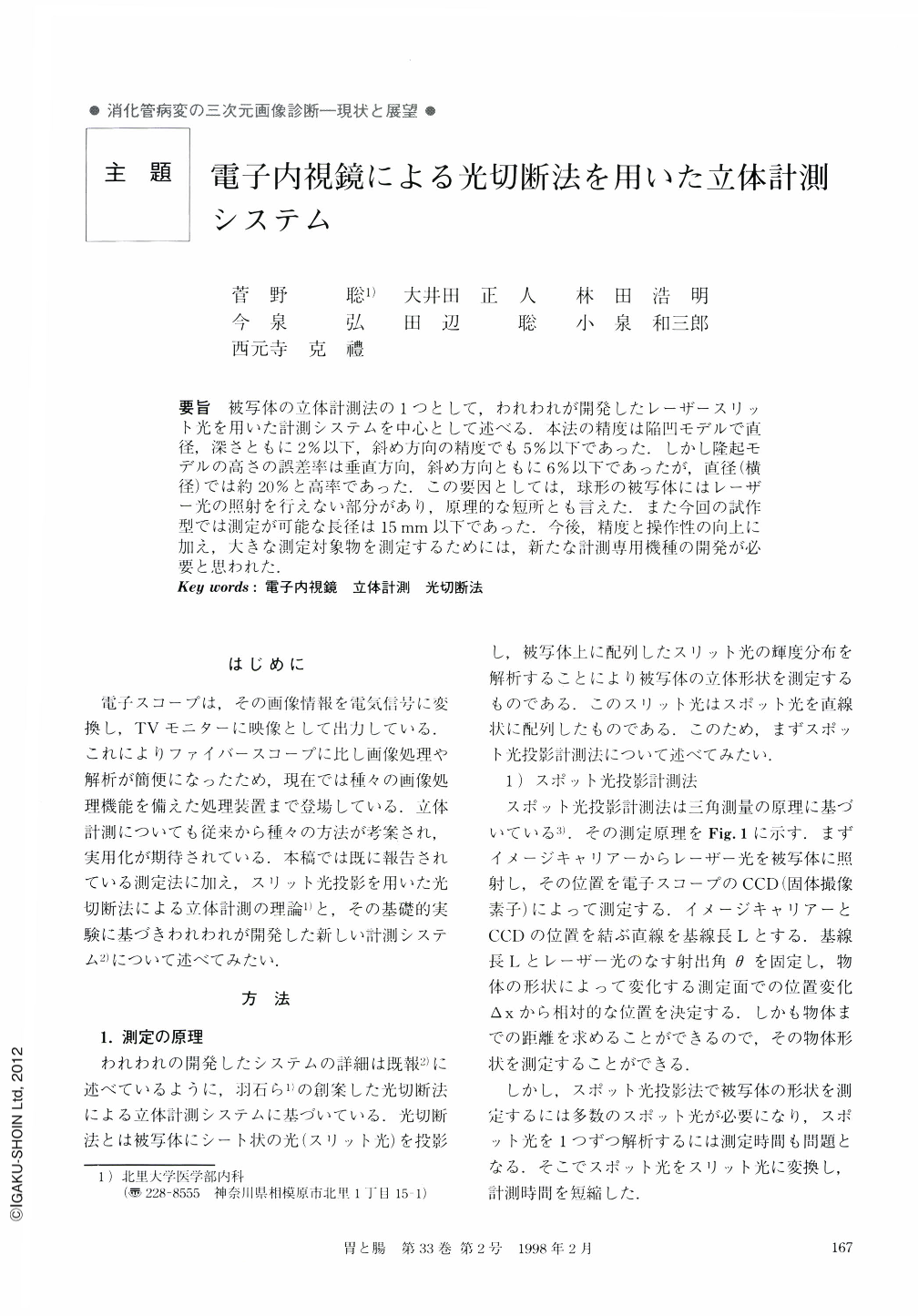
要旨 被写体の立体計測法の1つとして,われわれが開発したレーザースリット光を用いた計測システムを中心として述べる.本法の精度は陥凹モデルで直径,深さともに2%以下,斜め方向の精度でも5%以下であった.しかし隆起モデルの高さの誤差率は垂直方向,斜め方向ともに6%以下であったが,直径(横径)では約20%と高率であった.この要因としては,球形の被写体にはレーザー光の照射を行えない部分があり,原理的な短所とも言えた.また今回の試作型では測定が可能な長径は15mm以下であった.今後,精度と操作性の向上に加え,大きな測定対象物を測定するためには,新たな計測専用機種の開発が必要と思われた.
We experimented with a development of an electronic endoscopic system for three-dimensional measurement using slit beam projection. The materials used in three basic experiments included a Video system (CV200, Olympus), an electronic endoscopy (GIF type Q200, Olympus), reconstructed EVIP 230 for image processing. Slit laser beam was projected from an angiofiberscope to the measured objects, and the beam intensity distribution was measured three-dimensionally with image processing. It took about tree seconds for image processing. A protruding model and a depressed model were prepared, and the precision of measurement was studied from perpendicular (90°) and oblique (-70°~+110°) directions. We used a sphere type as a protruding model. The mean error rate of the depressed model was 3.1%. The mean error rate for the height of the protruding model was 5.2%, but the mean error rate for the diameter of the protruding model was 20.7%. The mean error rate for the diameter of the protruding model was higher than the ratio for the height of the protruding model. Apparently because the slit laser beam was not projected on the back surface of the spherical model, and measurement was not possible. We performed measurements of the shape of a human gastric ulcer, and we succeeded in almost accurate measurement of the shape of the ulcer. We concluded that clinical application of this method is feasible, but it is necessary for higher precision to design an electronic endoscopic system exclusively for three-dimensional measurements.

Copyright © 1998, Igaku-Shoin Ltd. All rights reserved.


