Japanese
English
- 有料閲覧
- Abstract 文献概要
- 1ページ目 Look Inside
- サイト内被引用 Cited by
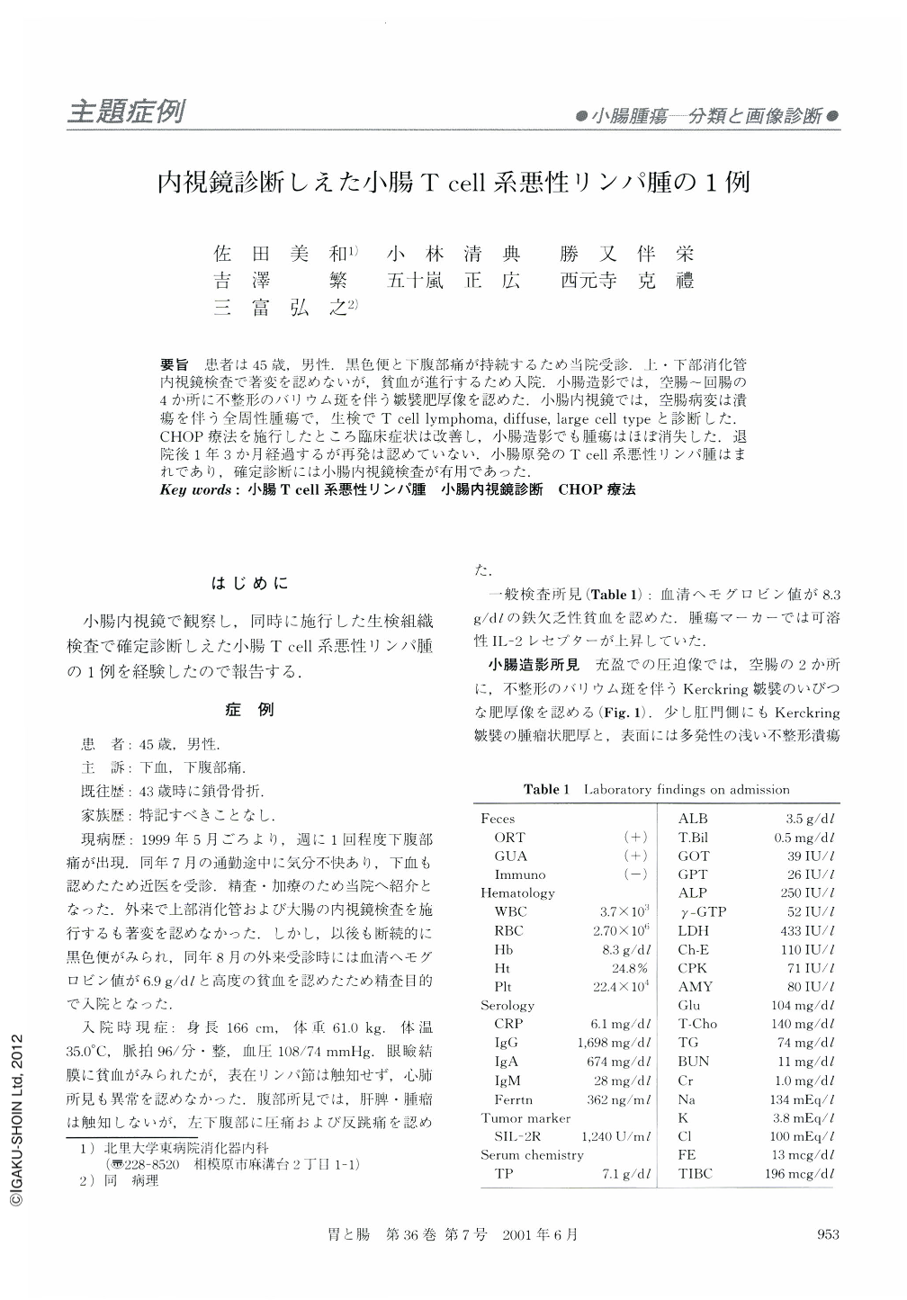
要旨 患者は45歳,男性.黒色便と下腹部痛が持続するため当院受診.上・下部消化管内視鏡検査で著変を認めないが,貧血が進行するため入院小腸造影では,空腸~回腸の4か所に不整形のバリウム斑を伴う皺襞肥厚像を認めた.小腸内視鏡では,空腸病変は潰瘍を伴う全周性腫瘍で,生検でT cell lymphoma,diffuse,large cell typeと診断した.CHOP療法を施行したところ臨床症状は改善し,小腸造影でも腫瘍はほぼ消失した.退院後1年3か月経過するが再発は認めていない.小腸原発のT cell系悪性リンパ腫はまれであり,確定診断には小腸内視鏡検査が有用であった.
A 45-year-old male was admitted to our hospital because of bloody stool and lower abdominal pain. Small intestinal radiography revealed diffuse thickness of Kerckring's folds with irregular-shaped ulcers in the middle and lower part of the jejunum and ileum. We observed the tumors by using a newly developed push-type small intestinal videoendoscope (SIF-Q240). Histological findings of the biopsied specimens showed that it was a non-Hodgkin's, diffuse, large-cell,T cell type of malignant lymphoma.
After three courses of CHOP therapy, the abdominal symptoms had diminished, and radiography revealed that small intestinal tumors comprised of malignant lymphoma had disappeared or decreased in size. The patient has had no recurrence of the disease in the 15 months since the last chemotherapy.
T-cell type of small intestinal malignant lymphoma is rare, and the newly developed videoendoscope (SIF-Q240) makes it easier diagnose malignant lymphoma in the upper part of the small intestine.

Copyright © 2001, Igaku-Shoin Ltd. All rights reserved.


