Japanese
English
- 有料閲覧
- Abstract 文献概要
- 1ページ目 Look Inside
- サイト内被引用 Cited by
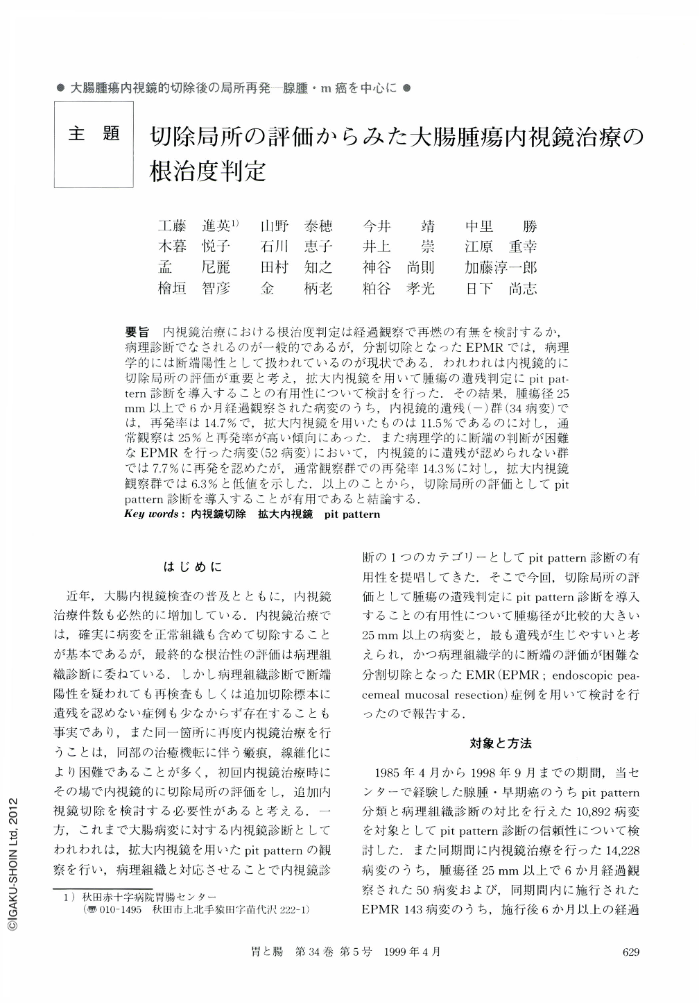
要旨 内視鏡治療における根治度判定は経過観察で再燃の有無を検討するか,病理診断でなされるのが一般的であるが,分割切除となったEPMRでは,病理学的には断端陽性として扱われているのが現状である.われわれは内視鏡的に切除局所の評価が重要と考え,拡大内視鏡を用いて腫瘍の遺残判定にpit pattern診断を導入することの有用性について検討を行った.その結果,腫瘍径25mm以上で6か月経過観察された病変のうち,内視鏡的遺残(-)群(34病変)では,再発率は14.7%で,拡大内視鏡を用いたものは11.5%であるのに対し,通常観察は25%と再発率が高い傾向にあった.また病理学的に断端の判断が困難なEPMRを行った病変(52病変)において,内視鏡的に遺残が認められない群では7.7%に再発を認めたが,通常観察群での再発率14.3%に対し,拡大内視鏡観察群では6.3%と低値を示した.以上のことから,切除局所の評価としてpit pattern診断を導入することが有用であると結論する.
It is generally known that complete resection by endoscopic therapy has been made on the basis of confirmation that the margin of the resected specimen contains normal glands in pathology (stated, “the margin of the resected specimen is negative”). But occasionally, we encounter cases of pathological incomplete resection which have been followed up clinically for a long time and have not resulted in recurrence. We suggest that there is a difference between pathological diagnosis and clinical diagnosis in cases of complete resection, and that in vivo estimation of the margin after endoscopic resection is important. Incidentally, we studied the surface structure of colorectal neoplasms (pit pattern) and by using magnifying endoscopy we are able to assert that pit pattern is a helpful diagnostic clue indicating the probability of recurrence or non-recurrence. This study was carried out to determine the therapeutic efficacy of using magnifying endoscopy in endoscopic resection, especially endoscopic piecemeal mucosal resection (EPMR). We did this because we were aware that EPMR illustrated many problems in diagnosing residual lesions. During the period from April 1985 to June 1998 we have encountered 14,228 endoscopic resections, and 50 cases which were over 25 mm in size and which we observed six months after resection. We also treated 143 cases using EPMR in the same period and analyzed 52 cases which could be sufficiently evaluated in respect to recurrence and residue for over six months in this study. When, by using magnifying endoscopy in vivo, we were able to observe only normal gland pits (type Ⅰ pit patterns) in the margin of the resection, we recognized the resection as clinically complete.
For lesions over 25 mm in size, pathology indicated that there would be recurrence in 14.7% of the cases. However, by using magnifying endoscopy in vivo as explained above, recurrence was predicted in only 11.5% of the cases.
In the case of EPMR the rate of recurrence was 7.7% in total.
By using ordinary endoscopy, the rate of recurrence was 14.3%, but, relying on in vivo magnifying endoscopy, the rate of recurrence was reduced to 6.3%.
In coclusion, it is stated that the clinical results of using magnifying endoscopy were investigated and its degree of effectiveness was shown to be excellent.

Copyright © 1999, Igaku-Shoin Ltd. All rights reserved.


