Japanese
English
- 有料閲覧
- Abstract 文献概要
- 1ページ目 Look Inside
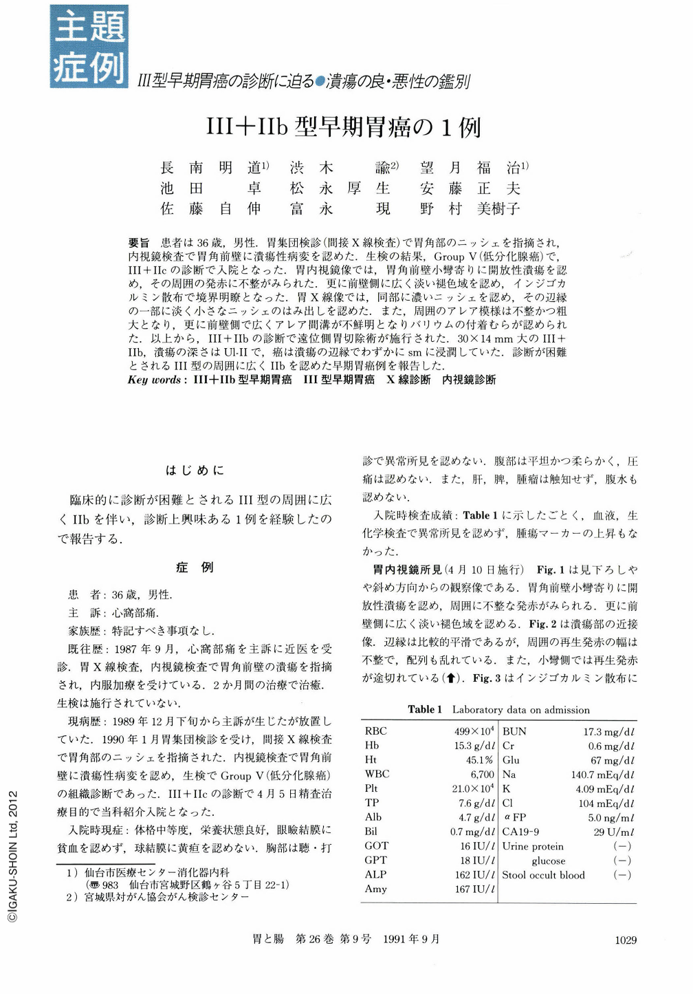
要旨 患者は36歳,男性.胃集団検診(間接X線検査)で胃角部のニッシェを指摘され,内視鏡検査で胃角前壁に潰瘍性病変を認めた.生検の結果,Group V(低分化腺癌)で,Ⅲ+Ⅱcの診断で入院となった.胃内視鏡像では,胃角前壁小彎寄りに開放性潰瘍を認め,その周囲の発赤に不整がみられた.更に前壁側に広く淡い褪色域を認め,インジゴカルミン散布で境界明瞭となった.胃X線像では,同部に濃いニッシェを認め,その辺縁の一部に淡く小さなニッシェのはみ出しを認めた.また,周囲のアレア模様は不整かつ粗大となり,更に前壁側で広くアレア間溝が不鮮明となりバリウムの付着むらが認められた.以上から,Ⅲ+Ⅱbの診断で遠位側胃切除術が施行された.30×14mm大のⅢ+Ⅱb,潰瘍の深さはUl-Ⅱで,癌は潰瘍の辺縁でわずかにsmに浸潤していた.診断が困難とされるⅢ型の周囲に広くⅡbを認めた早期胃癌例を報告した.
During a mass survey of the stomach, a 36-year-old male was found to have a niche at th egastric angle. Endoscopic examination was performed and an ulceration was detected at the anterior wall of the same region. Biopsy specimens which were obtained from this lesion showed it was a poorly differentiated adenocarcinoma. The patient was admitted to our hospital. Endoscopically, there was an ulceration surrounded by irregular redness at the gastric angle. Wide-spread light discoloration was shown at the anterior side of this lesion. Radiographically, a small and slight barium fleck next to the thick niche was shown surrounded by a rough and irregular area. And the pattern of the area on the anterior side of this lesion was coarse and unclear. We diagnosed the lesion as being Ⅲ+Ⅱb type early gastric cancer. Distal gastrectomy was performed. In the resected specimen, there was Ⅲ type gastric cancer, but we couldn't identify the Ⅱb part. Pathologically, the depth of the ulcer was Ul-Ⅱ, and cancer cell had invaded as far as the upper part of the submucosal layer at the margin of the ulcer.

Copyright © 1991, Igaku-Shoin Ltd. All rights reserved.


