Japanese
English
症例
術前3D-CTにより中葉肺静脈の破格を把握し安全に胸腔鏡下右下葉切除を施行しえた1例
Thoracoscopic Right Lower Lobectomy Safely Performed after the Identification of Anomalous Middle Pulmonary Vein by Preoperative Three-dimensional Computed Tomography Angiography:Report of a Case
守尾 篤
1
,
斎藤 雄一
2
,
今清水 恒太
3
,
鈴木 健司
3
Atsushi Morio
1
,
Yuichi Saito
2
,
Kota Imashimizu
3
,
Kenji Suzuki
3
1小張総合病院呼吸器外科
2帝京大学呼吸器外科
3順天堂大学呼吸器外科
1Department of Thoracic Surgery, Kobari General Hospital
キーワード:
肺癌
,
肺静脈
,
破格
Keyword:
lung cancer
,
pulmonary vein
,
anatomical anomaly
pp.239-241
発行日 2023年3月1日
Published Date 2023/3/1
DOI https://doi.org/10.15106/j_kyobu76_239
- 有料閲覧
- Abstract 文献概要
- 1ページ目 Look Inside
- 参考文献 Reference
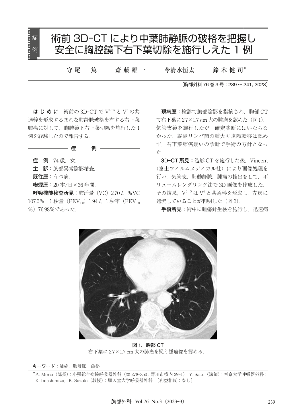
はじめに 術前の3D-CTでV4+5とV6の共通幹を形成するまれな肺静脈破格を有する右下葉肺癌に対して,胸腔鏡下右下葉切除を施行した1例を経験したので報告する.
A complete thoracoscopic right lower lobectomy was successfully performed for a 74-year-old woman with an anomalous right middle lobe pulmonary vein, forming a common trunk of V4+5 and V6. Preoperative three-dimensional computed tomography was useful to identify the vascular anomaly and contributed to perform safe surgery under the thoracoscopy.

© Nankodo Co., Ltd., 2023


