Japanese
English
- 有料閲覧
- Abstract 文献概要
- 1ページ目 Look Inside
- 参考文献 Reference
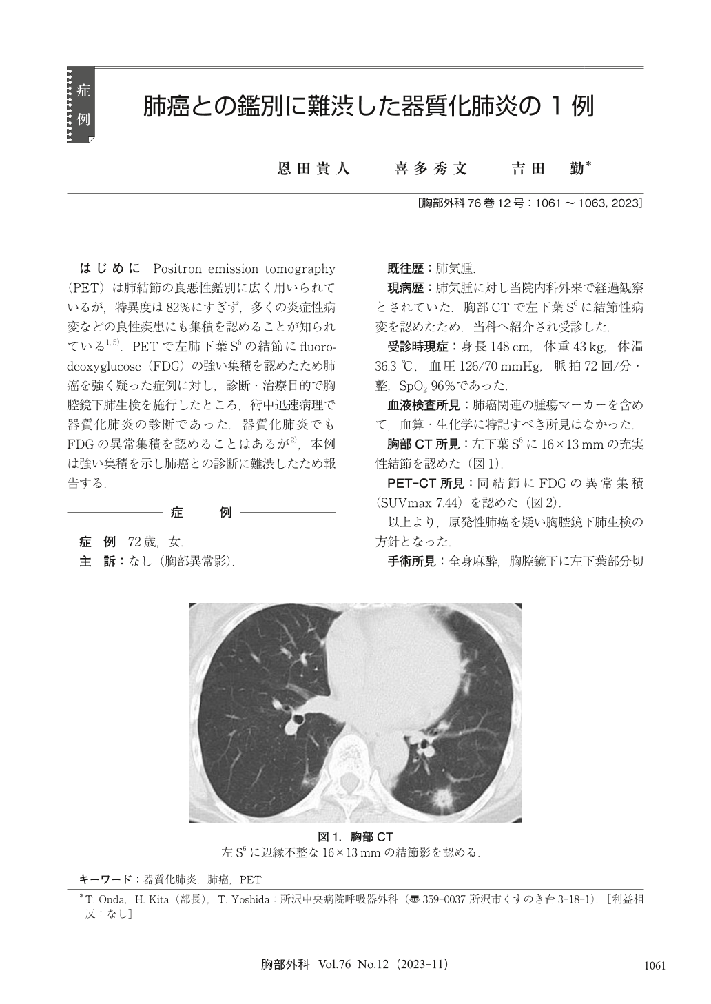
はじめに Positron emission tomography(PET)は肺結節の良悪性鑑別に広く用いられているが,特異度は82%にすぎず,多くの炎症性病変などの良性疾患にも集積を認めることが知られている1,5).PETで左肺下葉S6の結節にfluorodeoxyglucose(FDG)の強い集積を認めたため肺癌を強く疑った症例に対し,診断・治療目的で胸腔鏡下肺生検を施行したところ,術中迅速病理で器質化肺炎の診断であった.器質化肺炎でもFDGの異常集積を認めることはあるが2),本例は強い集積を示し肺癌との診断に難渋したため報告する.
A 72-year-old woman underwent a close examination because of chest computed tomography (CT) scan revealed a nodule in the left lower lobe of the lung. Positron emission tomography (PET) showed strong accumulation of fluorodeoxyglucose (FDG) in the lesion. Since lung cancer was strongly suspected, video-assisted thoracoscopic lung biopsy was performed. The lesion was diagnosed as organizing pneumonia by pathology. PET is widely used to distinguish between benign and malignant lung nodules, but FDG accumulation can also be seen in benign diseases such as inflammatory lesions. Abnormal accumulation can also be seen in organizing pneumonia, but strong FDG accumulation such as in this case is relatively rare, and it was difficult to distinguish it from lung cancer.

© Nankodo Co., Ltd., 2023


