Japanese
English
- 有料閲覧
- Abstract 文献概要
- 1ページ目 Look Inside
I.はじめに
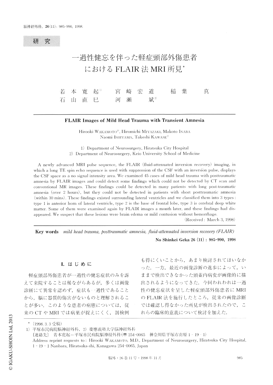
軽症頭部外傷患者が一過性の健忘症状のみを訴えて来院することは稀ながらあるが,多くは画像診断にて異常を認めず,症状も一過性であることから,脳に器質的傷害がないものと理解されることが多い.このような患者の病態については,従来のCTやMRIでは病巣が捉えにくく,剖検例も得にくいことから,あまり検討されてはいなかった.一方,最近の画像診断の進歩によって,いままで検出できなかった頭蓋内病変が画像的に描出されるようになってきた.今回われわれは一過性の健忘症状を呈した軽症頭部外傷患者にMRIのFLAIR法を施行したところ,従来の画像診断では確認し得なかった所見が検出されたので,これらの臨床的意義について検討を加えた.
A newly advanced MRI pulse sequence, the FLAIR (fluid-attenuated inversion recovery) imaging, in which a long TE spin echo sequence is used with suppression of the CSF with an inversion pulse, displays the CSF space as a no signal intensity area. We examined 45 cases of mild head trauma with posttraumatic amnesia by FLAIR images and could detect some findings which could not be detected by CT scan and conventional MR images. These findings could be detected in many patients with long post-traumatic amnesia (over 2 hours), but they could not be detected in patients with short posttraumatic amnesia (within 30 mins). These findings existed surrounding lateral ventricles and we classified them into 3 types :type 1 is anterior horn of lateral ventricle, type 2 is the base of frontal lobe, type 3 is cerebral deep white matter. Some of them were examined again by FLAIR images a month later, and these findings had dis-appeared, We suspect that these lesions were brain edema or mild contusion without hemorrhage.

Copyright © 1998, Igaku-Shoin Ltd. All rights reserved.


