Japanese
English
- 有料閲覧
- Abstract 文献概要
- 1ページ目 Look Inside
I.はじめに
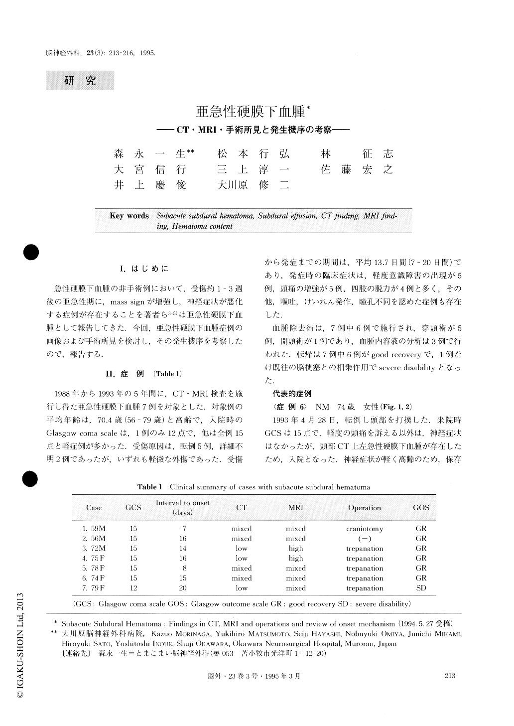
急性硬膜下血腫の非手術例において,受傷約1-3週後の亜急性期に,mass signが増強し,神経症状が悪化する症例が存在することを著者ら3-5)は亜急性硬膜下血腫として報告してきた.今回,亜急性硬膜下血腫症例の画像および手術所見を検討し,その発生機序を考察したので,報告する.
Subacute subdural hematoma was investigated in terms of findings in CT, MRI and operations and of onset mechanism.
The subjects were 7 cases of subacute subdural hematoma in which CT and MRI were performed dur-ing the past 5 year period. Subacute subdural hemato-ma here was defined as a nonoperated case with acute subdural hematoma, accompanied by subacute ex-acerbation 1 - 3 weeks after head trauma. The time from the injury to the onset averaged 13.7 days. CT re-vealed mixed density in 4 cases, low density in 3 cases and cerebral atrophy in all cases, with increasing mass sign due to the enlarged low density area. MRI re-vealed mixed intensity in 5 cases and high intensity in 2 cases, with increasing mass sign due to the enlarged high intensity area. Operation disclosed the outer mem-brane of the hematoma only in 1 case, but the inne, membrane could not be identified in any case. Analysi of the hematoma contents showed a low hemoglobin concentrations and a high level of methemoglobin. Lack of outer membrane in cases with subacute subdu-ral hematoma suggests that this is a different disease entity from chronic subdural hematoma. It is surmised that subacute subdural hematoma is the result of sub-dural effusion in the subacute stage, because, judging from the findings of CT and MRI each performed ovei time, cerebrospinal fluid is considered accountable for the increase in the mass sign.

Copyright © 1995, Igaku-Shoin Ltd. All rights reserved.


